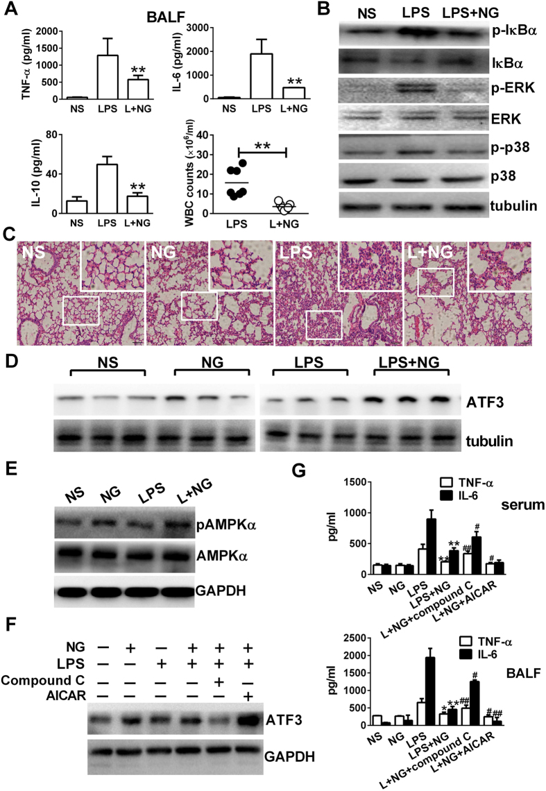
Figure 7. Naringenin upregulates ATF3 expression in lung tissues of LPS-challenged mice, which is AMPK dependent and required for limiting proinflammatory reactions.
(A,B) Detection of cytokines and cell counts in BALF and Western blot assays for signalling molecules in murine lung tissues. Mice were injected with NS, LPS or LPS with NG. BALF or lung tissues were collected 12 h after LPS injection. TNF-α, IL-6 and IL-10 levels and WBC counts (n = 3) in the BALF were measured (A). Protein levels of p-IκBα, IκBα, ERK, pERK, p-p38, p38 and tubulin were detected by western blot (B). (C–E) Detection of histological changes, ATF3 expression and AMPK activation in murine lung tissues. Mice were injected with NS, NG, LPS or LPS with NG, and lung tissues were collected 12 h after injection. Histopathological changes (C) were observed, and the protein levels of ATF3 (D) or pAMPKα and AMPKα (E) were detected by western blot. Uncropped images are presented in Supplementary Figure S8C,D. (F,G) Effects of the co-injection of AMPK modulators on the levels of ATF3 and proinflammatory cytokines. Mice were injected with NS, NG, LPS, LPS plus NG, LPS plus NG and compound C (1 mg/kg) or LPS plus naringenin and AICAR (100 mg/kg). Blood, lung tissues and BALF were collected 12 h after injection. ATF3 expression in the lung tissues were detected by western blot (F). TNF-α and IL-6 levels in the serum or in the BALF (G) were detected by ELISA (n = 3). *P < 0.05, **P < 0.01 vs LPS; #P < 0.05; ##P < 0.01 vs LPS+NG. Naringenin is abbreviated as NG. Broncho-alveolar lavage fluid is abbreviated as BALF. The doses of LPS and NG were both 10 mg/kg unless indicated.

