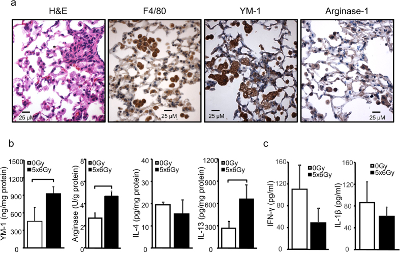
Figure 2. Type 2 inflammation in irradiated mouse lung.
Eight-10 week old female c57BL6/NcR mice were exposed to 5 daily fractions of 6 Gy (5 × 6 Gy) of thoracic irradiation. (a) Sections of lung tissue collected at 16 weeks after irradiation or no irradiation (0 Gy) were immunostained for F4/80, YM-1, and Arginase-1. (b) Lung tissue (n = 5 mice) was homogenized and YM-1, IL-13, and IL-4 levels were measured with ELISA. Arginase-1 activity was also assessed. (c) Levels of interferon-γ and IL-1β in BAL fluid were measured with ELISA. Columns: mean, error bars: +SD, brackets: p < 0.05: Student’s t-test.

