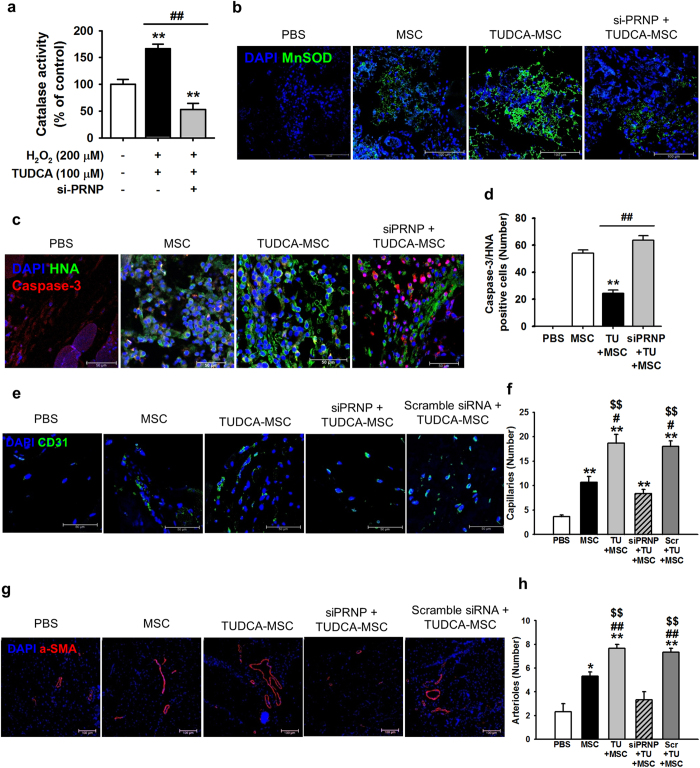
Figure 6. TUDCA-treated MSCs enhance functional recovery in murine hindlimb ischemia.
(a) In oxidative stress conditions (treatment with H2O2), TUDCA-mediated catalase activity in MSCs was assessed by determining the expression levels of PrPC. Values represent the mean ± SEM. **P < 0.01 vs. untreated MSCs in non-oxidative condition, ##P < 0.01 vs. TUDCA-treated MSCs pretreated with PRNP-specific siRNA under oxidative stress conditions. (b) At postoperative day 1, immunofluorescence staining for MnSOD (green) was performed in the ischemic-injured tissues of each group. Scale bar = 100 μm. (c) At postoperative day 3, apoptosis of transplanted MSCs was investigated by immunofluorescence staining for HNA (green) and cleaved caspase-3 (red). Scale bar = 50 μm. (d) Apoptotic transplanted MSCs were quantified based on the number of HNA and PCNA double-positive cells. Values represent the mean ± SEM. **P < 0.01 vs. transplantation of MSCs, ##P < 0.01 vs. transplantation of TUDCA-treated MSCs. (e–g) At postoperative day 28, capillary density and arteriole density were assessed by immunofluorescence staining for CD31 (green; e) and α-SMA (Red; g), respectively. Scale bar = 50 μm, 100 μm. Capillary density (f) and arteriole density (h) were quantified as the number of CD31 and α-SMA positive cells, respectively. Values represent the mean ± SEM. *P < 0.05 and **P < 0.01 vs. injection of PBS, #P < 0.05 and ##P < 0.01 vs. transplanted MSCs, $$P < 0.01 vs. transplantation of TUDCA-treated MSCs pretreated with PRNP-specific siRNA.

