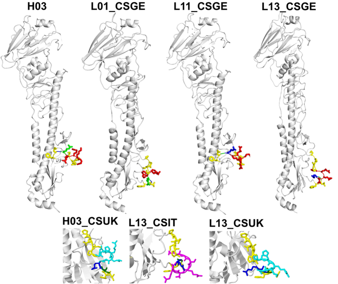
Figure 7. Molecular modelling of the HA protein.
Predicted tertiary structure of the HA proteins of selected recombinants with pCS. Variable cleavage site motifs are illustrated: cleavage site between arginine (blue: R) and glycine (G: green), monobasic CS is depicted in yellow sticks; pCSGE in red sticks; pCSIT in magenta sticks; pCSUK in cyan sticks.

