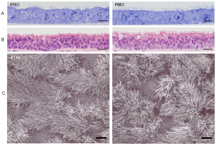
Figure 1. Morphological examination of porcine well-differentiated airway epithelial cell cultures.
(A) PTEC and PBEC cultures were grown under ALI conditions for more than 4 weeks. The semi-thin sections followed by toluidine blue staining were performed. (B) Epithelia from porcine trachea and primary bronchus were collected, followed by histological sectioning and H&E staining for the morphological comparison. The histological examination was evaluated by light microscopy and the representative histological sections (40x magnification) are shown. (C) The micrograph of the scanning electron microscopy illustrates the apical surface of PTEC and PBEC. The ciliated epithelial cells are the predominant cell type. Scale bars, 20 μm (A,B), 5 μm (C).

