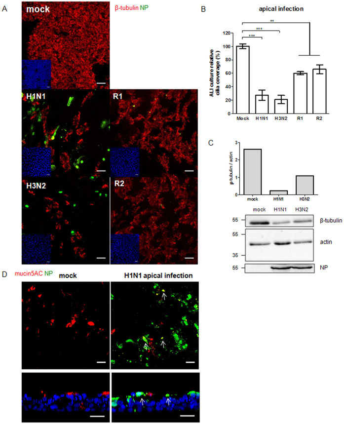
Figure 6. Immunofluorescent staining of porcine well-differentiated airway epithelial cells at 8 days post infection.
WdPBECs were inoculated with IAV from the apical side at an MOI of 0.25 and fixed at 8 dpi. (A) PBEC cultures were stained for viral nucleoprotein (green) and cilia (red). (B) Quantification of the ciliated area at 8 dpi. Results are shown as percentages (means ± SEM) compared to mock-infected cultures. For each infection, six PBECs from three independent donors were measured, and three fields per culture were evaluated as technical replicates. (C) Western blot analysis of β-tubulin expression level in PBECs after swIAV infection. The relative expression level of β-tubulin was normalized to actin expression. The viral NP could be detected in the infected culture. (D) Immunofluorescent staining for viral nucleoprotein (green) and mucin (red). The nuclei were stained by DAPI (blue) (A and D). The arrows show co-localization. Scale bars, 25 μm.

