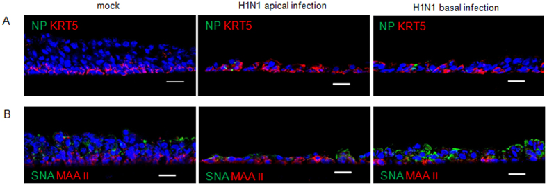
Figure 9. Sialic acid expression on PBEC after swIAV infection.
WdPBECs were inoculated by swIAV H1N1 from the apical (middle panels) or basolateral (right panels) side at an MOI of 0.25. Cryosections were prepared at 8 dpi. (A) Immunofluorescent staining for KRT 5 (red, basal cells) and viral nucleoprotein (green). (B) Immunofluorescent staining to detect α2,3- and α2,6-linked sialic acid using MAA II and SNA lectins, respectively. The nuclei were stained by DAPI (blue) (A&B). Scale bars: 25 μm.

