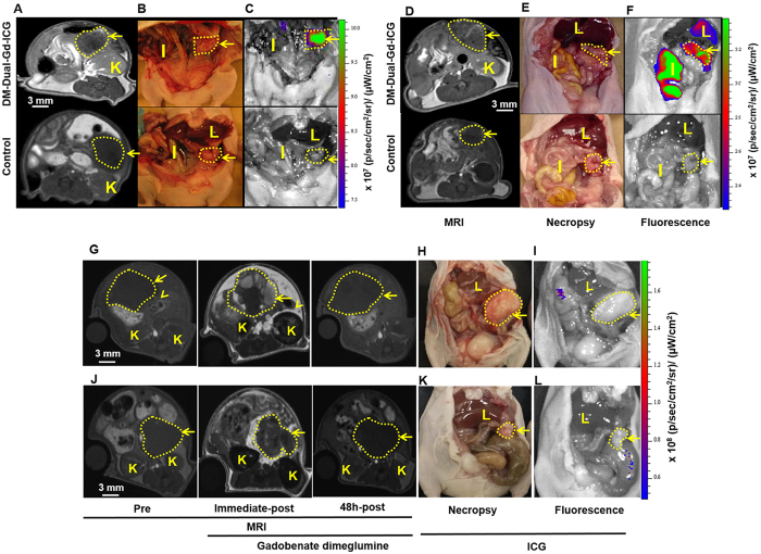
Figure 3. In vivo MR imaging and NIRF imaging shows that DM-Dual-Gd-ICG results in enhancement of HeyA8 tumors 2 days later.
Representative axial 2D-FSPGR MR images of mice injected IV with DM-Dual-Gd-ICG (top) or vehicle (bottom) (A). Representative coronal necropsy (B) and coronal open abdomen near infrared fluorescence images (C) of a nude mice. In vivo MR imaging and NIR imaging shows that DM-Dual-Gd-ICG liposomes results in enhancement of OVCAR-3 tumors 2 days later. Representative axial 2D-FSPGR MR images of mice injected IV with DM-Dual-Gd-ICG (top) or vehicle (bottom) (D). Representative coronal necropsy (E) and coronal open abdomen near infrared fluorescence images (F) of a nude mice. Representative MR and NIR imaging show that free ICG or gadobenate dimeglumine do not result in increased signal in HeyA8 tumors 2 days after IV injection. Representative axial 2D-FSPGR MR images pre, immediate-post and 2 days after IV injection of gadobenate dimeglumine (G). Representative coronal necropsy (H) and coronal open abdomen near infrared fluorescence images (I) of nude mice. Representative MR and NIR imaging show that free ICG or gadobenate dimeglumine do not result in increased signal in OVCAR3 tumors 2 days after IV injection. Representative axial 2D-FSPGR MR images pre, immediate-post and 2 days-post after IV injection of gadobenate dimeglumine (J). Representative coronal necropsy (K) and coronal open abdomen near infrared flouresence images (L) of a nude mice. Arrow, tumor; I, intestine; K, kidney; L, Liver.

