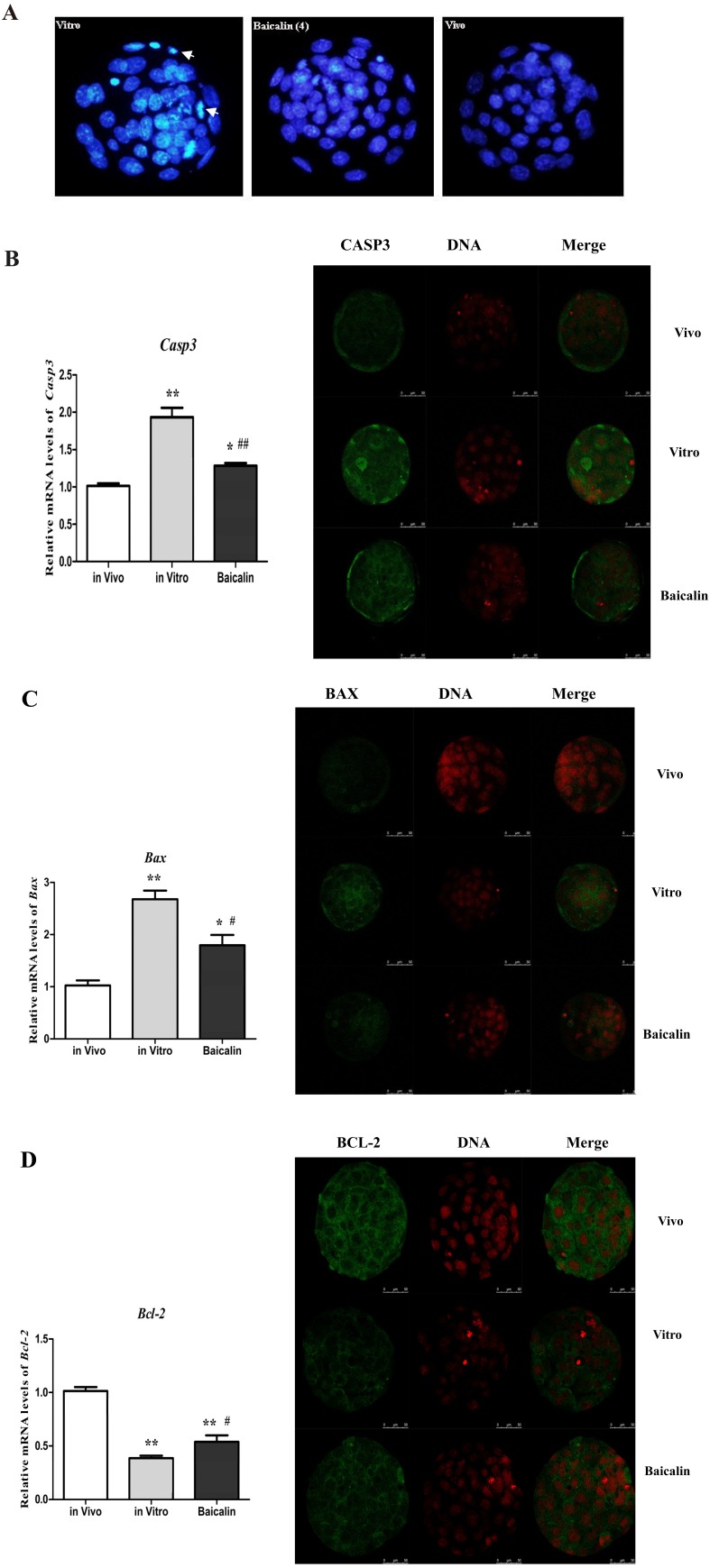
Fig. 4.
Effect of baicalin on cellular apoptosis in mouse embryos. The nuclei of blastocysts (n = 30) were stained with Hoechst 33342 to examine cell apoptosis (Bar = 100 μm). Blastocysts from the in vitro (left), baicalin-treated (middle), and in vivo (right) groups showing higher degree of apoptosis of nuclei for in vitro group (white arrows) than the baicalin treated or in vivo group (A). Relative mRNA and protein expression levels of Caspase-3 (B), BAX (C) and BCL-2 (D), respectively, in mouse blastocysts (n = 30) from the in vitro, baicalin treated and in vivo groups. * P < 0.05 vs. in vivo control group and ** P < 0.01 vs. in vivo control group; # P < 0.05 vs. IVC group and ## P < 0.01 vs. IVC group.

