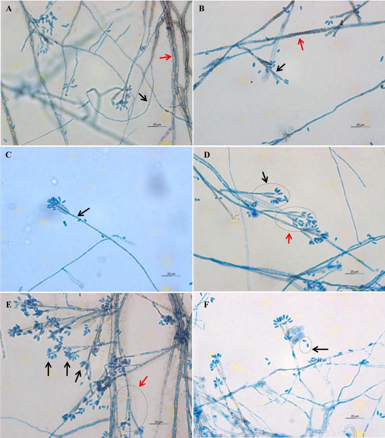
Figure 4. Microscopic morphology of D. eschscholtzii isolates.
(A) Thin-walled and hyaline septate hyphae (black arrow), thick-walled and melanized septate hyphae (red arrow). (B) Pigmented exudates on hyphae surface (red arrow), additional branch grew from the conidiogenous regions (black arrow). (C) Mononematous conidiophore (black arrow) with conidiogenous cells arising from its terminus. (D) Conidiophore with dichotomous branching, with two (black arrow) to three (red arrow) conidiogenous cells arising from each terminus. (E) Conidia were produced holoblastically in sympodial sequence on the terminus of the conidiogenous cells (black arrows), conidiophore with trichotomous branching pattern (red arrow). (E) Ellipsoid conidia with attenuated base (black arrow). (400× magnification, bars 20 µm).

