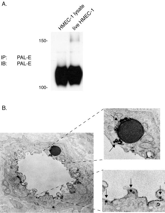
FIG. 5.
PAL-E-reactive vimentin is expressed on the luminal endothelial cell surface in association with endothelial vesicles. A. The PAL-E antigen detected on the surfaces of live HMEC-1 cells is identical to the one detected in total cell lysate. Immunoblot analysis was performed with PAL-E antibody on protein immunoprecipitated from total HMEC-1 cell lysate, using PAL-E-conjugated beads (left lane), and on protein immunoprecipitated from the surface of live HMEC-1 cells exposed to unconjugated PAL-E antibody and washed prior to lysis (“live cell” IP, right lane). Note the precipitation of identical 120-kDa proteins under both conditions. B. PAL-E-reactive vimentin is expressed in association with endothelial vesicles along the luminal cell membrane. Indirect immunoelectron microscopy was performed after binding with PAL-E antibody to a frozen human skin tissue section. Strong binding is observed around vesicles on the luminal cell membrane (lower right) and around a trapped erythrocyte (upper right) (magnification, ×76,000).

