Figure 1.

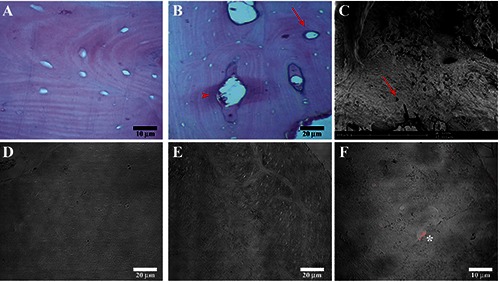
Compound panel of FFB grade 0 images obtained by hematoxylin-eosin, scanning electron microscopy and immunofluorescence techniques. A) Hematoxylin-eosin images show bone tissue characterized by empty osteocyte lacunae and also the presence of empty Volkmann’s (head arrow) and Haversian canals (arrow) (B). C) Scanning electron microscopy image shows FFB grade 0 characterized by empty osteocyte lacunae (red arrow) and absence of cellular components. Immunofluorescence reactions images show the absence of fluorescence for RANKR (D) and osteocalcin (E); in F) it is also possible to observe the presence of VEGF fluorescence pattern within some osteocyte lacunae (asterix).
