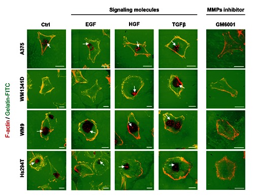
Figure 4.

Microscopical analysis of effects of EGF, HGF or TGFβ on melanoma cell lines’ invadopodia formation and extracellular matrix degradation potential. Melanoma cells were seeded in medium containing only 20% FBS (control) or containing 20% FBS with addition of EGF, HGF or TGFβ onto coverslips coated with fluorescently labeled gelatin. As a negative control the cells were treated with medium containing 20% FBS and 25 μM GM6001, a pan-inhibitor of matrix metalloproteases. 16 h later the cells were fixed and stained with Alexa Fluor 568-labeled phalloidin. Invadopodia are indicated with white arrows. Gelatin degradation is visualized by lack of a green fluorescence signal. Scale bar: 20 μm.
