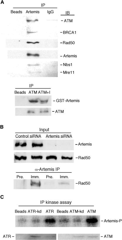
FIG. 4.
Artemis interacts with checkpoint proteins and is phosphorylated by ATM and ATR. (A, top blot) Artemis antibodies coimmunoprecipitate ATM, BRCA1, Rad50, Nbs1, and Mre11 from HeLa extracts. Protein A-Sepharose beads were used as a control. IP, immunoprecipitation; IgG, nonspecific immune serum. Immunoblotting (IB) was performed using antibodies to the indicated proteins. (A, lower blot) Reciprocal coimmunoprecipitation of Artemis by ATM antibodies. ATM+I, incubation of the extracts with DNase I prior to coimmunoprecipitation. (B) Depletion of Artemis in HeLa cells by transfection of siRNA eliminates the coimmunoprecipitation of Rad50 by Artemis antibodies. α-Artemis IP, immunoprecipitation with anti-Artemis antibodies; Pre., preimmune serum; Imm., Artemis antiserum. (C) Immunoprecipitation (IP) kinase assay shows that Artemis was phosphorylated by both ATM and ATR but by kinase-dead (kd) variants of the enzymes. The top shows an autoradiogram of Artemis phosphorylation by ATM or ATR. The bottom shows immunoblots of immunoprecipitated kinases used in the assays.

