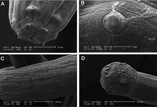
Figure 2.

Scanning electron micrographs of Eustrongylides spp. fourth-stage larva. A) Cephalic end: oral orifice, surrounded by cephalic papillae, scale bar = 50 μm; B) cephalic papilla of external circle, high magnification, scale bar = 5 μm; C) middle part of the worm showing transverse striations of the cuticle, scale bar = 200 μm; D) rounded posterior end of the larva, scale bar = 200 μm.
