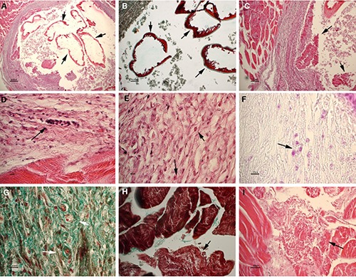
Figure 3.

Larva encapsulated in host muscle. A-B) Transverse section of the parasite showing intact cuticular layers (arrows). A) Haematoxylin-eosin (HE), scale bar = 200 μm; B) masson trichrome stain (MT), scale bar 100= pm. C) The arrows point out degenerating and necrotic larvae comprised inside the lumen of the capsule. HE, scale bar = 100 μm. DF) Magnification of the connective capsule surrounding the larva. Arrows point out lymphocytes (D, HE, scale bar = 10 μm), eosinophils (E, HE, scale bar = 10 μm) and macrophages (F, PAS, scale bar = 20 μm) infiltrating the capsule. G) Neoformed microvessels are evident in the wall of the capsule. MT, scale bar = 20 μm. H-I) Degenerating and necrotic muscle fibers surrounding the capsule. H) MT, scale bar=30 μm. I) HE, scale bar = 30 μm.
