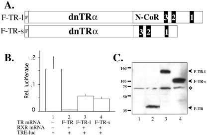
FIG. 1.
The fusion of the RID of N-CoR to a dominant negative TR leads to partial derepression. (A) Diagram of TR fusion proteins. The fusion constructs F-TR-l and F-TR-s include a long and short fragment of Xenopus N-CoR containing the RID. The black boxes labeled 3, 2, and 1 represent the corepressor nuclear receptor binding motifs (I/LXXI/VI) of N-CoR that directly bind TR (21). The dominant negative Xenopus TRα (dnTRα) part of the fusion protein corresponds to Xenopus TRα without the terminal helix 12 (3, 44). Both fusion proteins also contain an N-terminal FLAG tag. (B) The fusion proteins have reduced abilities to repress T3 target genes in the frog oocyte transcription system. The plasmids for the dual luciferase reporter system, where the firefly luciferase was driven by the T3-inducible Xenopus TRβA promoter (TRE-luc) and the Renilla luciferase was driven by the T3-independent TK promoter, were injected into the oocyte nucleus. The mRNAs for RXR and FLAG-tagged wild-type TRα (F-TR), F-TR-l, or F-TR-s were injected into the cytoplasm. After overnight incubation in the absence of T3, the oocytes were harvested for luciferase assay, and the relative expression of the reporter firefly luciferase over the control Renilla luciferase was determined. The experiment was repeated with similar results. (C) Western blot showing the expression of the receptor proteins in the frog oocyte. Protein extracts from luciferase assay from lanes 1 to 4 as shown in panel B were separated by SDS-PAGE, and FLAG-tagged wild-type and mutant TRs were detected by anti-FLAG Western blotting. Molecular weight markers are shown, and the asterisk indicates a cross-reacting band.

