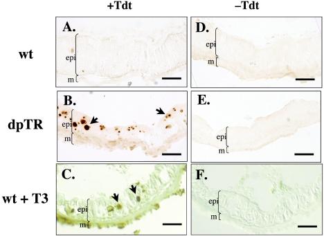
FIG. 5.
Expression of F-dpTR causes larval intestinal epithelial cells to undergo apoptosis. Wild-type and F-dpTR transgenic tadpoles were reared in methimazole to block metamorphosis and then heat shocked. A TUNEL assay to detect apoptotic cells was carried out on cross sections of intestine after 4 days of heat shock. For comparison, wild-type tadpoles were treated with 5 nM T3 for 3 days before the TUNEL assay. Wild-type tadpoles without T3 treatment showed no TUNEL labeling (A), whereas the intestine of F-dpTR transgenic and T3-treated tadpoles had many stained cells, indicating cell death (B and C, black arrows). Lack of staining in the sections performed without terminal TdT revealed the specificity of the reaction (D to F). Brackets indicate boundaries of muscles (m) and larval intestinal epithelium (epi) facing the intestinal lumen (not marked is a thin layer of connective tissue present between the muscles and epithelium). The color differences among sections were due to photography but do not affect the conclusion. This experiment was repeated three times with similar results. Bars, 25 μm.

