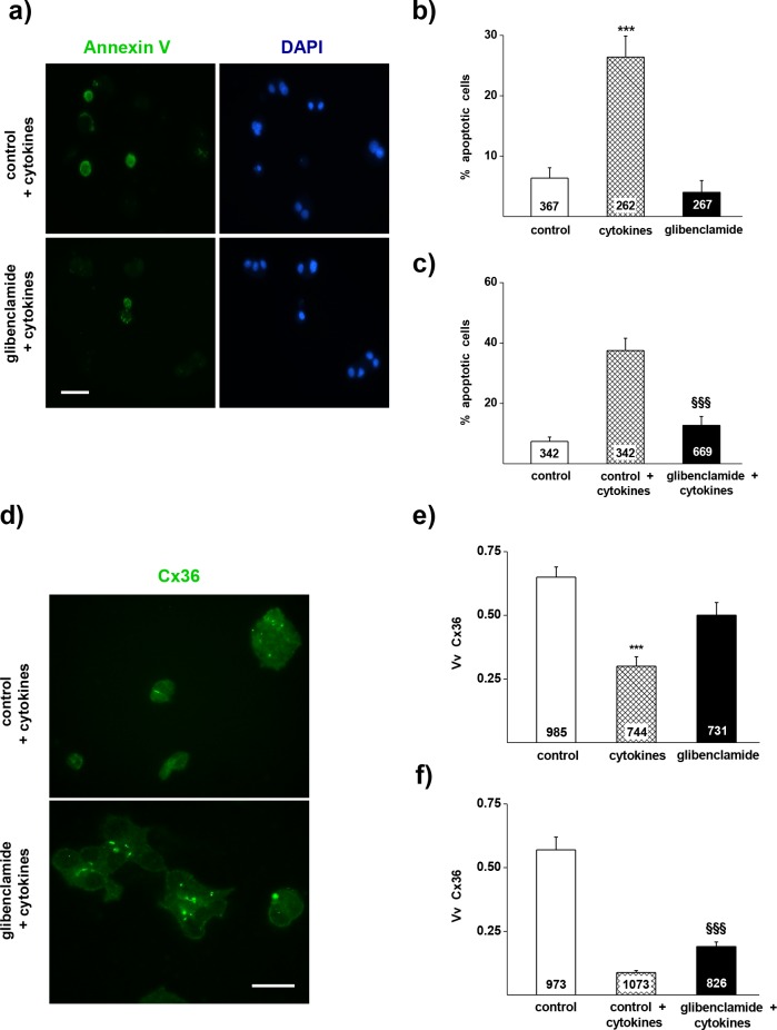
Fig 1. Glibenclamide reduces the apoptosis and the loss of CVx36 in MIN6 cells exposed to Th1 cytokines.
a) Apoptosis of MIN6 cells was evaluated by the proportion of Annexin V-stained cells (green) out of the total number of cells screened (identified by the blue DAPI staining of nuclei). Bar, 25 μm. b) A 18h exposure of MIN6 cells to a mix of IL-1β, IFN γ and TNFα induced a significant increase in the proportion of Annexin V-labelled cells (hatched bar), compared to the level observed in controls cultured in the absence of cytokines (open bar). Such an increase was not observed in cells exposed to 10 μM glibenclamide (solid bar). c) Comparable effects were seen when MIN6 cells were exposed to 10 μM glibenclamide 6 h before starting the exposure to the cytokines. d) Expression of Cx36 was evaluated by immunostaining, using an antibody that detects the small gap junction plaques made by the concentration of Cx36 within the membrane (yellow-green). Bar, 5 μm. e) A 18h exposure of MIN6 cells to IL-1β, IFN γ and TNFα (hatched bar) reduced the volume density of immunolabeled Cx36 under the control levels (open bar). Such a decrease was not observed in cells exposed to 10 μM glibenclamide (solid bar). f). Comparable effects were seen when MIN6 cells were exposed to 10 μM glibenclamide 6 h before starting the exposure to the cytokines. Data are mean + SEM of 3 independent experiments. Numbers within bars indicate the total number of cells scored in each group. *** p < 0.001 vs control group;. §§§ p < 0.001 vs control + cytokines group, as tested by one-way ANOVA.

