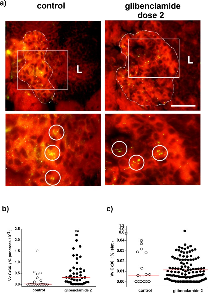
Fig 4. Glibenclamide treatment preserves Cx36 expression in beta cells.
a) At the end of the 25 wk experiment, immunostaining reveals the spotted distribution of Cx36 (yellow) in the few residual islets of control, untreated mice and in the more numerous islets of the glibenclamide dose 2-treated mice. The enlargements of the field outlined in the pictures show that Cx36 formed larger and more numerous spots (some are circled) in the islets of the latter animals. The dot line outlines the limit between an islet and its surrounding insulitis mantle (L). Bar, 40 μm in the pictures, 20 μm in the enlargements. The red background staining is due to the Evans blue counterstain. b) The volume density of Cx36 was significantly lower in the whole pancreas of untreated controls than in that of the glibenclamide-treated mice, due to the much larger number of islets found in the latter animals. c) In contrast, no significant difference in the volume density of Cx36 was reached in the residual islets of the 2 groups of mice that we compared, possibly because few surviving islets could be scored in the pancreas of the untreated NOD mice. ** p< 0.01 vs control group, as tested by Mann-Whitney and median tests. Median values are shown by the red lines.

