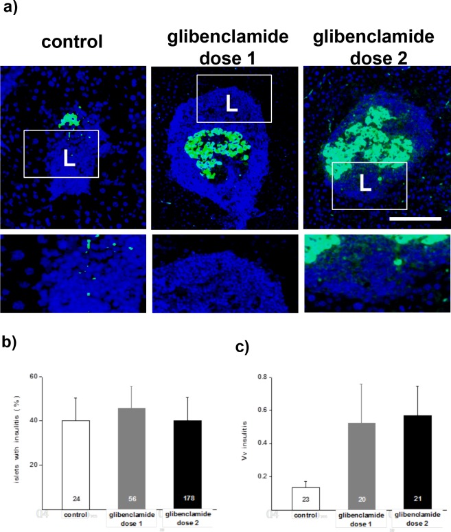
Fig 5. Islets of untreated control and glibenclamide-treated NOD mice display insulitis.
a) Few beta cells are immunostained for insulin (green) in the islets of untreated controls, used for morphometry, whereas many more beta cells are seen in the islets of the animals treated with glibenclamide. The blue DAPI staining outlines the insulitis mantle (L). Lower panels are enlargements of the fields outlined in the pictures to visualize the dense packing of the immune cells, mostly lymphocytes that formed the insulitis mantle. Bar, 100 μm in the pictures, 40 μm in the enlargements. b) All mice featured a similar percentage of islets with insulitis. c) However, a higher volume density of insulitis per unit pancreas volume was found after the glibenclamide treatments, due to the much larger number of islets surviving under these conditions. Data are mean + SEM values of the number of islets (b) or pancreas sections (c) indicated within the bars. * p< 0.05, ** p< 0.01, *** p< 0.001 vs control group, as tested by ANOVA.

