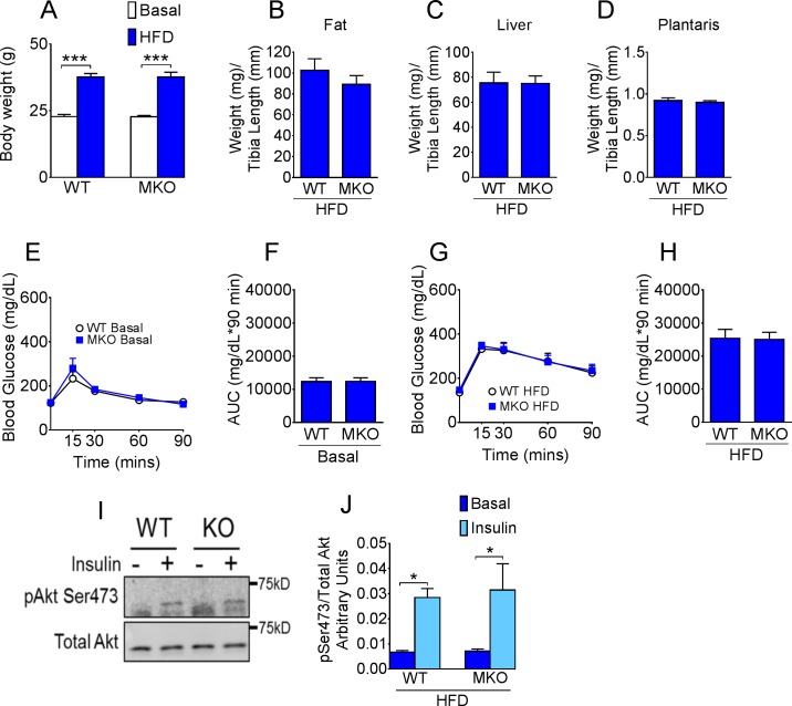
Fig 3. CypD deletion in skeletal muscle does not affect whole body glucose homeostasis.
(A) Body weight at basal levels and following 11 wk of HFD; (B) Epididymal fat; (C) liver and (D) plantaris muscle tissue weight normalized to tibia length in HFD fed mice; (E) Blood glucose following i.p. injection of glucose in normal chow fed mice and (F) area under the glucose curve; (G) Blood glucose following i.p. injection of glucose in HFD fed mice and (H) area under the glucose curve; (I) Representative western blot and (J) quantification of phosphorylated (S473) and total Akt in plantaris muscle of HFD fed WT and MKO mice before and 10 min after i.p. injection of insulin. ***p < 0.001 basal vs. HFD; *p < 0.05 basal vs. insulin; n = 10–12.

