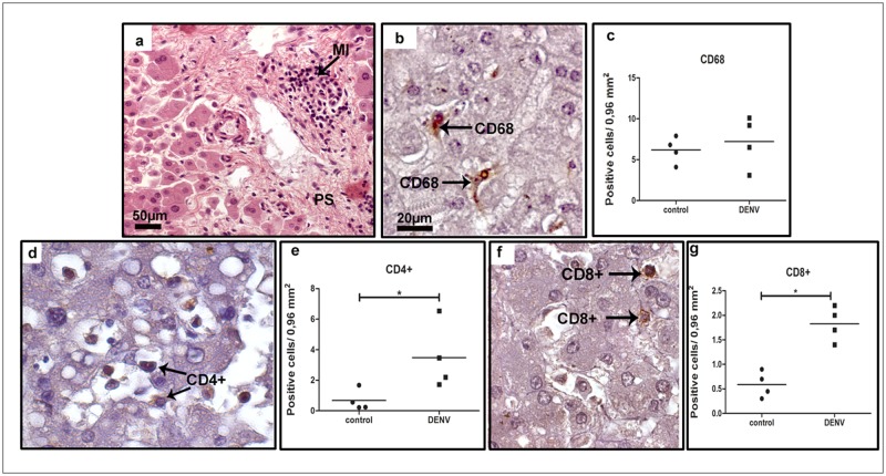
Fig 1. Characterization of cell subpopulations in liver tissues from DENV-3 fatal cases.
Sections were stained with (a) H.E. or (b, d, f) incubated with specific antibodies in immunohistochemical assays. (a) Liver of a representative dengue case showing diffuse mononuclear infiltrates around the portal space; (b) detection of hyperplasic Kupffer cells (CD68+) observed mainly in sinusoidal capillaries of the dengue cases; (c) quantification of CD68+ cells in dengue cases and controls (non-dengue cases); (d) detection of CD4+ cells manly in portal space of the dengue cases; (e) quantification of CD4+ cells in controls and dengue cases; (f) CD8+ cells detected mainly in portal space; (g) quantification of CD8+ cells. MI—mononuclear cell infiltrates. Asterisks indicate differences that are statistically significant between dengue cases and control groups, (*p < 0.05). Staining controls are shown in S1 Fig.

