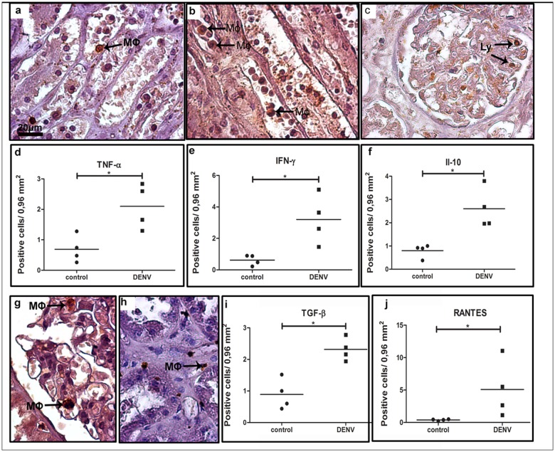
Fig 6. Cytokine-producing cells in renal tissues of dengue cases.
(a) TNF-α detected in monocytes located in blood vessels; (b) Production of INF-γ observed in circulating macrophages and lymphocytes located in the interstitial space; (c) IL-10 found in endothelial cells of glomerulus; (g) TGF-β production by lymphocytes present mainly inside renal glomeruli; (h) RANTES detected in macrophages and lymphocytes in the interstitial renal space; (d-f, i and j) quantification of the number of cells expressing the above cytokines. Macrophages (Mϕ); Lymphocytes (Ly). Asterisks indicate differences that are statistically significant between analyzed groups (*p < 0.05). Staining controls are shown in S3 Fig.

