
Figure 1.
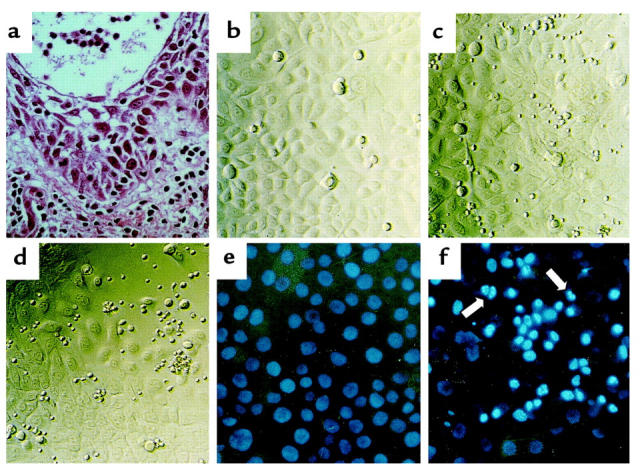
(a) Representative histologic findings of acute eczematous dermatitis. A dense subepidermal inflammatory infiltrate and marked epidermal acantholytic and spongiotic changes progress to vesicle formation. Hematoxylin/eosin staining. ×400. (b–d) Signs of KC injury after coculture with autologous T cells. Photomicrographs from 96-well plates with an inverted microscope equipped with phase contrast. ×200. (b) Intact monolayer of cultured third-passage primary human KCs. The KCs are relatively uniform in size and morphology. (c) Intact monolayer of KCs after 3 days in coculture with unstimulated autologous CD45RO+ T cells. (d) Partly destroyed monolayer of KCs after 3 days in coculture with autologous CD45RO+ T cells stimulated with anti-CD2, anti-CD3, and anti-CD28 mAb’s. (e and f) Induction of KC apoptosis in vitro. Identification of apoptotic nuclei with HOECHST staining. ×200. (e) Intact monolayer of KCs after 3 days of coculture in Transwell plates with unstimulated CD45RO+ T cells. (f) Induction of KC apoptosis after coculture in Transwell plates with stimulated CD45RO+ T cells. Note bright, condensed nuclei and nuclear fragmentation (arrows), signs of KC apoptosis.