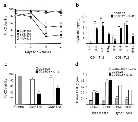
Figure 4.
Induction of KC apoptosis by type 1 and type 2 T cells. (a) Type 1 but not type 2 T cells mediated KC death. Coculture of primary human KCs and heterologous differentiated type 1 and type 2 T cells. AP < 0.05. (b) Cytokine content of differentiated and stimulated type 2 T cells as determined by ELISA. CD4+ T helper 2 (Th2) and CD8+ T cytotoxic 2 (Tc2) cells were stimulated with anti-CD2, anti-CD3, and anti-CD28 mAb’s or with a combination of anti-CD2, anti-CD3, and anti-CD28 mAb’s and IL-12. AP < 0.05. (c) Coculture of primary human KCs and heterologous differentiated type 2 T cells. KC viability was measured by ethidium bromide exclusion and flow cytometry at day 3. Control, KCs alone. CD4+ Th2 cells and CD8+ Tc2 cells were stimulated with anti-CD2, anti-CD3, and anti-CD28 mAb’s or with a combination of anti-CD2, anti-CD3, and anti-CD28 mAb’s and IL-12. AP < 0.05. (d) Levels of soluble FasL in T-cell supernatants as determined by ELISA. CD4+ Th1/Th2 and CD8+ Tc1/Tc2 cells were stimulated with anti-CD2, anti-CD3, and anti-CD28 mAb’s or with a combination of anti-CD2, anti-CD3, and anti-CD28 mAb’s and IL-12. Results shown represent three (a, c) to five (b, d) experiments and are shown as mean ± SD from triplicate cultures.

