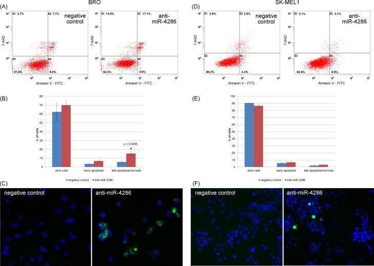
Fig 5. Effect of anti-miR-4286 on melanoma cell apoptosis.
(A) Flow cytometry showed a significant increase in apoptotic BRO melanoma cells after miR-4286 inhibition. (B) The percentage of viable, early apoptotic, late apoptotic/necrotic BRO cells. (C) Fluorescent microscopy of alive and apoptotic BRO cells: viable cell nuclei are colored blue, and the cytoplasm of apoptotic caspase-3+/7+ cells is colored green. *—significant difference compared to the apoptosis rate of negative control cells. (D) Flow cytometry revealed no changes in apoptosis of SK-MEL-1 cells after miR-4286 inhibition. (E) The percentage of viable, early apoptotic, late apoptotic/necrotic SK-MEL-1 cells. (F) Fluorescent microscopy of alive and apoptotic SK-MEL-1 cells: viable cell nuclei are colored blue, and the cytoplasm of caspase-3+/7+ apoptotic cells is colored green.

