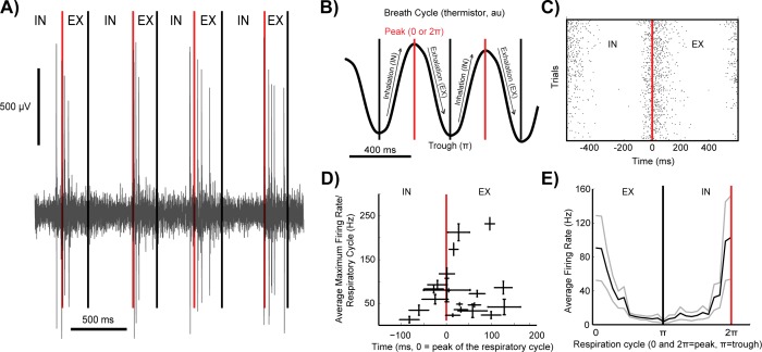
Fig 1. MTCs exhibit burst firing at the transition from inhalation to exhalation.
(A) Spontaneous burst firing in an in vivo extracellular single unit recording from the mitral cell layer. (B) A thermistor recording of respiratory cycle activity. Black arrows denote periods of inhalation and exhalation. Red and black lines highlight the peaks and troughs, respectively, of each thermistor respiratory cycle. (C) A raster plot of spikes across the respiratory cycle of the neuron in panel A. Red line at 0 ms is the time of the peak of the respiratory cycle. (D) Average maximum firing rates (Hz) relative to the thermistor peak voltage (n = 21 single unit recordings, 20 mice) and (E) across the respiratory cycle (cycle angles 0 to π to 2π, radians) shown in black, plus and minus standard errors in grey (n = 10 single unit recordings, 8 mice).

