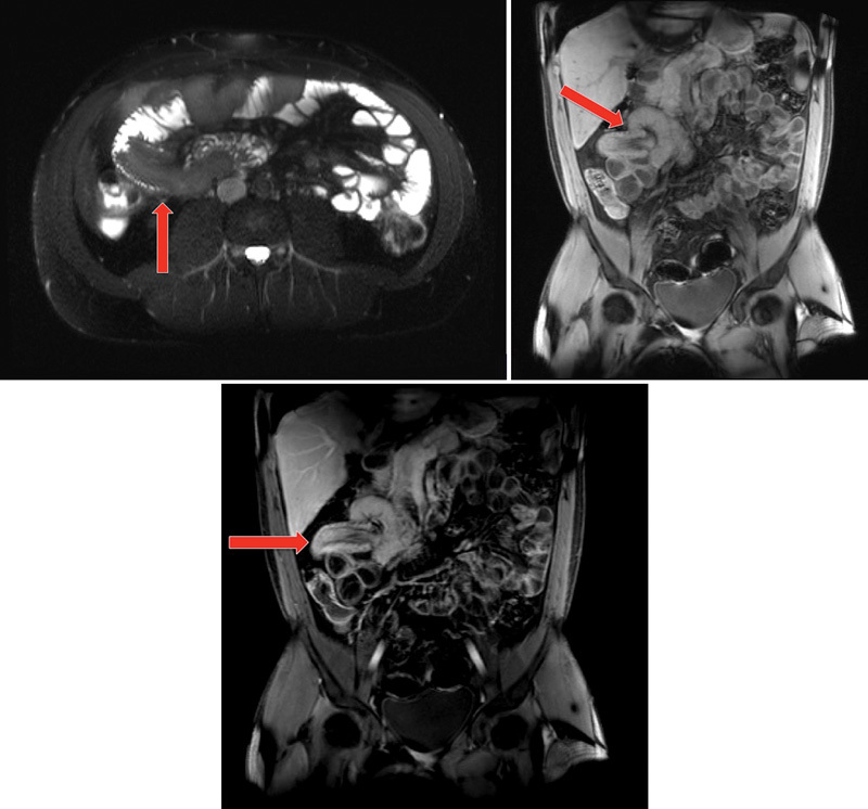
Fig. 5.

Magnetic resonance enterography (MRE). Left panel demonstrates a sausage-shaped filling defect in the right hemi-abdomen; the middle (contrast enhanced) and right (postcontrast) panels demonstrates the invagination. (Images courtesy of Dr. Nancy McNulty, MD.)
