



Abstract
Paradoxical puborectalis contraction (PPC) and increased perineal descent (IPD) are subclasses of obstructive defecation. Often these conditions coexist, which can make the evaluation, workup, and treatment difficult. After a thorough history and examination, workup begins with utilization of proven diagnostic modalities such as cinedefecography and anal manometry. Advancements in technology have increased the surgeon's diagnostic armamentarium. Biofeedback and pelvic floor therapy have proven efficacy for both conditions as first-line treatment. In circumstances where PPC is refractory to biofeedback therapy, botulinum toxin injection is recommended. Historically, pelvic floor repair has been met with suboptimal results. In IPD, surgical therapy now is directed toward the potentially attendant abnormalities such as rectoanal intussusception and rectal prolapse. When these associated abnormalities are not present, an ostomy should be considered in patients with IPD as well as medically refractory PPC.
Keywords: obstructive defecation syndrome, paradoxical puborectalis contraction, descending perineum syndrome, increased perineal descent, dyssynergic defecation, biofeedback
Chronic functional constipation affects 2 to 30% of individuals in Western countries.1 The Rome III criteria guide physicians in classifying functional constipation; however, two-thirds of the time the different subtypes of functional constipation coexist,2 3 4 5 and symptoms alone cannot differentiate between the pathologic subgroups.6 As opposed to disorders in colonic motility, the subtype of obstructive defecation in its inherent context describes the inability to evacuate contents from the rectum. Paradoxical puborectalis muscle contraction (PPC) and increased perineal descent (IPD) (or more commonly termed descending perineum syndrome) are two conditions in the spectra of obstructive defecation syndrome (ODS). In 2008, Clinics of Colon and Rectal Surgery published a review article in the workup of these two subclasses of ODS by Wexner and Landmann. This current article serves as a review and update on the diagnostic and treatment options available.
Paradoxical Puborectalis Contraction
In the literature, several different terms have been used to describe the paradoxical anal contraction rather than the normal relaxation during attempted defecation. Glia et al7 and Wexner et al8 discussed the role of PPC in ODS and the role of biofeedback. In patients with a normal anal sphincter, the puborectalis muscle relaxes and the anorectal angle widens, thus straightening the rectum and permitting the passage of stool during defecation. However, in patients with outlet obstruction from PPC, a paradoxical contraction of the puborectalis muscle occurs, resulting in an increase in the anorectal angle, thus prohibiting the normal passage of stool. Some authors argue that multiple terms misrepresent the physiologic abnormality. Recently, a panel of experts concluded that pathologic paradoxical anal contraction during attempted defecation was most represented by the term dyssynergic defecation (DD).9 Some authors use the terms DD and PPC interchangeably; however, it is important to recognize that some types of dyssynergia (i.e., type 3) do not involve PPC. The four types of DD will be discussed later.
Epidemiology
About one-third of patients with chronic constipation have an evacuation disorder, and of that one-third, most have DD.10
Pathophysiology
Under normal circumstances, when a patient attempts defecation, a rise in rectal pressure ensues with a synchronized voluntary relaxation of the external anal sphincter. Rao contends that the inability to perform this voluntary-coordinated maneuver is the chief pathophysiologic defect in patients suffering from obstructive defecation secondary to DD.11
History
Symptoms alone are poor predictors of pelvic floor dysfunction12; however, Rao reports in a prospective study of patients with DD and ODS that 85% of patients report excessive straining, 75% report feeling of incomplete evacuation, and 66% require digital maneuvers to facilitate defecation.13 Additionally, patients diagnosed with these defecation disorders display a variety of psychologic conditions.14
Physical
Digital rectal exam (DRE) is the most cost-effective tool available for the evaluation of patients suffering from ODSs including PPC. Orkin et al15 describe the digital rectal exam scoring system for assessing patients with paradoxical contraction of the anal sphincter complex, particularly assessing the anal resting and squeeze pressure. Their results correlated well with conventional anorectal manometry. Furthermore, DRE shows high sensitivity and positive predictive value in detecting dyssynergia of the anal sphincter compared with high-resolution anorectal manometry (HRARM). Soh et al emphasize that DRE should be utilized as a screening tool in workup of patients with symptoms of ODS and PPC and not a confirmatory test.16
Diagnostic Evaluation
Anorectal Manometry
Once physicians have a high suspicion for dyssynergia, anorectal manometry should be considered in the workup, most notably because manometry can exclude Hirschsprung disease by confirming the presence of the rectoanal inhibitory reflex.11 Rao also describes four types of dyssynergia that can be recognized by anorectal manometry. In short, the obstructive patterns are defined as follows: Type 1—paradoxical increase in the anal sphincter pressure in response to a driving force and rise in intrarectal pressure; Type 2—The patient is unable to manifest an adequate pushing force and will exhibit a paradoxical anal contraction; Type 3—the patient has the ability to manifest adequate pushing force and increase the intrarectal pressure but has less than 20% relaxation of resting anal sphincter pressure; and Type 4—the patient is unable to generate a sufficient driving force to increase intrarectal pressure and will manifest incomplete anal sphincter relaxation.11
Cinedefecography
Cinedefecography has been historically described as the gold standard for the evaluation and diagnosis of patients with dyssynergia.17 In the absence of other pelvic etiologies, cinedefecography can identify the paradoxical contraction rather than relaxation of the puborectalis muscle, the prolonged retention of contrast material, and the inability to evacuate the rectum.18 However, some authors challenge the clinical significance of contrast defecography due to overlap of abnormal findings such as rectocele, intussusception, and mucous prolapse. Piloni et al argue that these overlaps in findings in symptomatic patients and healthy controls question the clinical relevance of defecography.19 Because of this and other inherent deficiencies, some authors recommend using defecography only as an adjunct to clinical assessment for assessing DD.20 We often utilize defecography as an adjunct to anorectal manometry in evaluating patients for PPC.
Electromyography
Early on electromyography was utilized for assessment of the puborectalis. One study evaluated the importance of needle placement with external anal sphincter and puborectalis electromyography and evaluated the strain squeeze index in patients with constipation. After analyzing 164 patients versus 16 control subjects, it was concluded that quantification of puborectalis contraction and external anal sphincter with strain squeeze index of more than 50 differentiates patients in whom paradoxical activity may be the cause of constipation.21 Conversely, a retrospective study showed that electromyography has a low positive predictive value and a high negative predictive value for diagnosing PPC. It was recommended that electromyography alone is not an optimal test for diagnosing the presence of PPC.18
Balloon Expulsion Testing
An established criterion for diagnosing DD includes the inability to expel a 50-mL balloon after an established period of time. Some authors report the low negative predictive value of balloon testing and sensitivity and indicate that balloon expulsion was not an ideal screening test for DD in women with constipation.3 22 Conversely, Minguez et al had different results.23 In an effort to establish a method to exclude the possibility of pelvic dyssynergia in patients with constipation, Minguez et al prospectively studied two groups of patients with functional constipation. The control group consisted of patients with functional constipation without pelvic dyssynergia. The study group consisted of patients with functional constipation and pelvic dyssynergia that was previously diagnosed retrospectively by manometric and defecographic findings. Results of the study by Minguez et al showed that balloon expulsion test was pathologic in 21 of 24 patients with pelvic dyssynergia and the specificity and negative predictive value of the test for excluding pelvic dyssynergia were 89 and 97%, respectively. Minguez et al concluded that balloon expulsion is a useful screening procedure for patients with pelvic floor dyssynergia.23 Consensus data support that balloon expulsion test should be integrated with other anorectal exams such as anorectal manometry.24
High-Resolution Anorectal Manometry
HRARM differs from traditional manometry in that the HRARM makes use of more pressure sensors spaced at 1-cm intervals instead of 3- to 5-cm intervals with the intent of obtaining more detailed and reliable data. Kang et al argue that HRARM is likely to provide better physiological information with a statistically shorter measurement time. More evidence needs to be established, however.25
Treatment
The literature is well established on the initial treatment for patients with PPC. In addition to standard increased dietary fiber and fluid intake, patients should be instructed on timed toilet training and refraining from digital disimpaction.3 Patients not responding to standard treatment should be offered short-term biofeedback training. Several randomized controlled trials (RCTs) report that manometric-assisted pelvic relaxation, simulated defecation training, and pelvic floor therapy for DD are superior to standard treatments for constipation. Most patients experienced improved symptoms or even resolution of symptoms.26 27 28 Long-term data support the use of biofeedback for dyssynergia. In RCTs over a 1-year period, Rao reports that the number of complete spontaneous bowel movements per week improved compared with the standard therapy group. The dyssynergia pattern normalized, balloon expulsion time improved, and colonic transit time normalized only in the biofeedback group.29 Not only can biofeedback be effective in the long term, but data also show there is a significant impact on the improvement of quality of life (QOL) of patients suffering from DD.30 Consensus data now support Level I, Grade A recommendations for short-term and long-term biofeedback therapy in patients with dyssynergia.31 There are no accepted guidelines for optimal duration of therapy for biofeedback treatment. However, in cases where patients fail to initially respond to biofeedback, it is still reasonable to offer additional therapy sessions given its sometimes slow response rate and low risk nature.
Botox
Evidence suggests that Botox injection into the puborectalis muscle is an alternative or adjunct to biofeedback.32 In a prospective study of patients diagnosed with a nonrelaxing puborectalis, Shafik and El-Sibai reported that symptoms of straining at defecation disappeared and stool frequency normalized after injection of Botox into the external anal sphincter.33 Although short-term results (4.8 ± 1.4 months) are successful, patients often need reapplication. Khaikin and Wexner support the use of botulinum toxin injection in patients with PPC who have failed conservative treatment.34
Transanal Stimulation
In 2015, Cadeddu et al evaluated transanal electrostimulation (EST) that was designed to eliminate inappropriate contraction of the external anal sphincter during defecation attempts.35 Cadeddu et al evaluated in a RCT the coadministration of biofeedback plus transanal EST as the initial treatment for ODS. The study group undergoing EMG biofeedback plus transanal EST had a significant decrease in the mean Wexner score (16.7 ± 4 to 10 ± 3.5, p < 0.0102) and ODS (18.3 ± 5.5 to 5.7 ± 1.8, p < 0.0001). The QOL score also improved significantly from 61 ± 8.6 to 23 ± 4.8 (p < 0.0001). Conclusion of the study supported the used of combined biofeedback plus EST for initial treatment for patients with dyssynergia.
Surgical Treatment
Historically, surgical treatment of patients with DD has been scrutinized secondary to morbidity, especially incontinence, and the inability to produce adequate symptom relief. Surgical division of the puborectalis has been described for treatment of patients suffering from anismus, but despite initial satisfactory short-term results, patients report high rates of incontinence.36 37 However, in a 2009 small randomized study, Farid et al compared the results of bilateral partial division of puborectalis (PDPR) versus local botulinum toxin type A injection in patients with anismus. By dividing the inner half of the puborectalis muscle and its fibers attached to the rectal wall to widen the anorectal angle, these authors were able to achieve initial (100%) and long-term success (66.6%) in many.38 Minor incontinence was encountered in 2 of the 15 patients (13.3%). They concluded that bilateral PDPR is a promising method for patients with anismus. Long-term and larger studies need to be performed prior to endorsement of PDPR. It is still the practice of the senior author (L.G.) of this current review to offer a stoma to those patients refractory to all nonoperative attempts at therapy who still insist on operative intervention.
Increased Perineal Descent
The concept of studying the anorectal angle and measuring the distance of perineum descent was proposed by Parks et al.39 In this original article, they investigated patients with symptoms now recognized as obstructive defecation and calculated that 10% of patients with these symptoms experienced abnormal or increased descent of their perineum with defecation as measured by the pubococcygeal line, a line between the pubis and coccygeal ligament.
Pathophysiology
On defecography evaluation of normal individuals upon straining, the perineum can descend up to 3 cm from pubococcygeal line and the postdefecation reflex returns the pelvic floor to its normal anatomic position.39 Parks et al postulated that the underlying cause of IPD occurs in the setting of excessive straining against an outlet obstruction. If there is recurrent excessive straining, the pelvic floor muscle tone will weaken, the pelvic floor descends, and ultimately it becomes funnel shaped secondary to the stretching of the puborectalis and upper part of the sphincter muscles. After expulsion of the fecal bolus, normal individuals experience a postdefecation reflex and the pelvic floor and anorectal angle is restored. Because excessive straining is an inhibitor to pelvic muscle tone, repetitive straining will abolish the postdefecation reflex and eventually progress to permanent descent of the perineum. Further progression of the disease culminates with eventual external sphincter incompetence with resultant anal leakage. Early on, Kiff et al described severe neurogenic damage to the external anal sphincter muscle and its pudendal innervation.40 Damage to pudendal nerves can occur during perineal descent leading to denervation and weakness of the external anal sphincter muscle. Further weakness of the pelvic floor and straining eventually leads to prolapse of anterior rectal mucosa into the anal canal with eventual intussusception and possibly prolapse.39
Over the years, research has shown that IPD is not limited to the posterior compartment. Literature suggests that weakness of one compartment of the pelvic floor will increase the likelihood of dysfunction of additional or even all compartments of the pelvic floor.41 However, a comparison trial conducted by Soligo et al comparing constipated and nonconstipated women concluded that bowel dysfunction correlates exclusively with posterior pelvic compartment dysfunction.42 Even with this ongoing debate, Pucciani suggests that urogynecological structures are implicated and thus pelvic organ prolapse may coexist with other physical exam findings associated with IPD.43
History
Evidence suggests symptoms of IPD can be attributed to obstructed defecation, fecal incontinence, and urogynecological symptoms. This can manifest in varying degrees in relation to the disease stage.44 45
Pucciani describes an evolution of symptoms in IPD beginning with classic signs of ODS, such as excessive straining and occasionally manual maneuvers to facilitate evacuation of the rectum. This may progress to symptoms of fecal incontinence and fullness in the rectum, suggesting rectoanal intussusception. Occasionally, this can progress to full-thickness rectal prolapse. Attendant pudendal neuropathy and progressive increased descent portend external anal sphincter dysfunction and worsen the incontinence. Urological symptoms include urinary complaints and vaginal symptoms including sensation of a bulge.43
Physical
On digital rectal examination, the muscle tone may be decreased or normal. In most cases, the muscle tone is easily overcome by posterior traction with the examining finger. Characteristically during straining, the puborectalis muscle descends sharply and can no longer be felt as a separate bar constituting the anorectal ring. The upper anal canal becomes funnel shaped and the anterior rectal wall is pushed down on the examining finger.39
Diagnostic Evaluation
Early on, electromyography was used to evaluate IPD. However, increased usage of morphologic and physiologic tests throughout the years has improved physicians' ability to accurately diagnose IPD and improve diagnostic accuracy of attendant conditions.
Defecography
Defecography findings for IPD are represented by descent of the anorectal angle greater than 3 cm below the pubococcygeal line on straining.46 The anorectal angle may return very slowly to baseline or remain fixed.43 Owing to the ability to identify multiple abnormalities within different pelvic compartments, dynamic proctography and cystocolpoproctography (DCP) has largely replaced defecography alone as the workup of choice for IPD. DCP combines vaginal opacification and voiding cystography and defecography to visualize multiple compartments of the pelvis. One study supported the use of DCP and reported the significant benefit of DCP in changing the management of patients suffering from anorectal disorders up to 40% of the time.47 Conversely, Ahmad et al argue that without opacification of the bladder, only the posterior compartment can be visualized. They conclude that DCP should only be utilized for assessment of the posterior compartment and that DCP is unsuitable for evaluation of multicompartmental defects.48
MRI Defecography
MRI defecography has been increasingly utilized as a diagnostic tool for functional constipation, and previous grading scores for detection of pelvic floor abnormalities have been recognized.49 On MRI, the anorectal junction should not descend below the pubococcygeal line, so any descent below this classifies as IPD (Figs. 1 and 2). Recently, Piloni et al modified a previously described MR-based grading system for ODS.19 They concluded that even though cinedefecography was comparable to MR defecography regarding detection of common abnormalities including rectocele, intussusception, and prolapse, MR defecography offers a lack of ionizing radiation and more reproducible measurement of recognized parameters such as anorectal angle and increased pelvic organ descent on straining. Additionally, MR defecography was able to demonstrate occult anatomical defects affecting the pelvic diaphragm and concluded that these detected abnormalities impacted clinical decision making. The article maintained that the vast majority of clinicians who utilized the MR-based ODS classification system chose a different therapeutic option.
Fig. 1.
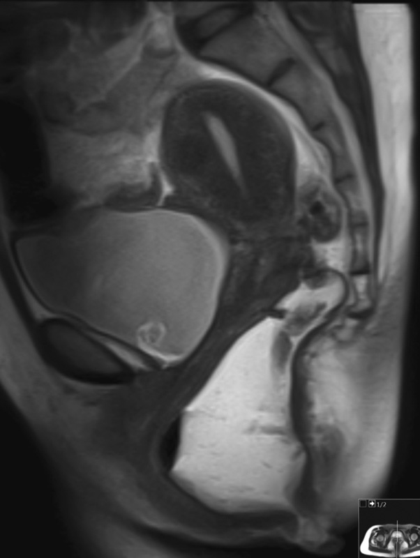
MRI defecography image showing significant descent of the rectum below the pubococcygeal line. (Images courtesy of Dr. Roopa Ram at the University of Arkansas for Medical Sciences.)
Fig. 2.
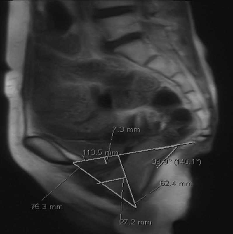
MRI defecography image showing increased perineal descent (6 cm) of the pelvic floor structures below the pubococcygeal line. (Images courtesy of Dr. Roopa Ram at the University of Arkansas for Medical Sciences.)
Transperineal Sonography
Standard transperineal sonography will likely be replaced by four-dimensional (4D) dynamic transperineal sonography. Standard transperineal sonography lacks the ability to assess defecation and micturition,43 but 4D dynamic transperineal ultrasound (DTP-US) provides real-time morphologic changes during pelvic floor contractions and allows for levator function to be assessed.50 Beer-Gabel et al compared DTP-US with defecation proctography and showed DTP-US was able to diagnose 71% of perineal descent, 75% of rectal prolapse, 92% of rectoceles, whereas three cases of perineal descent were missed by defecation proctography.51 An advantage of DTP-US is the visualization of all three pelvic compartments.
3D Endovaginal Ultrasound
O'Leary et al examined the posterior pelvic floor compartment with three-dimensional endovaginal ultrasound in patients already diagnosed with ODS. They measured anorectal angle and levator plate descent angle, and a multivariable logistic regressions analysis concluded that greater levator plate descent and widened anorectal angle are associated with ODS.52
3D High-Resolution Anorectal Manometry
In a comparative pilot study evaluating excessive perineal descent, Benezech et al compared 3D HRARM versus defecography.53 In 19 patients already diagnosed with IPD by defecography, anal canal pressures were calculated at rest and during straining with 3D HRARM. During straining, excessive perineal descent was defined as the downward movement of the anal high-pressure zone. With arrest of the straining effort, the high-pressure zone regained its initial position, thus indicating the probe had not moved. It was concluded that 3D HRARM can diagnose excessive perineal descent with the same degree of reliability as defecography.
Treatment
Biofeedback
Like PPC, biofeedback is attempted only in IPD after diet and lifestyle modifications have failed.34 Patients with IPD have shown significant improvement in their ODS score after biofeedback therapy.54 Patients with progression to fecal incontinence can anticipate improvement of the Wexner incontinence score with the addition of pelvic floor retraining and biofeedback therapy.55 Because of the associated urogynecological abnormalities, it is of benefit to seek a multidisciplinary approach including rehabilitative therapy.43 56 57 Rehabilitative therapy including pelviperineal kinesiotherapy aimed at improving muscular tone and function at the levator ani muscles should be involved in treatment algorithms. Kinesiotherapy can improve pelvic floor muscle tone and coordination and contribute to positive outcomes in anorectal surgery.56 57
Over recent years, there has been growing support for surgical treatment of patients with IPD resistant to pelvic floor retraining. Still, surgery is reserved for patients with reparable anatomical defects and other attendant pathologies.34 Historically, direct pelvic floor repair was met with suboptimal results. As such, surgeons are navigating away from pelvic floor surgery and focusing on treatments addressing surgically correctable concomitant rectal diseases, finding that surgical correction of these rectal diseases will indirectly improve the IPD.43
Rectal Intussusception (Internal Rectal Prolapse) and Rectocele
Nearly half of patients with rectal intussusception suffer from ODS.58 However, most of these patients benefit from dietary modification and biofeedback therapy. Often surgical therapy is primarily reserved for patients with coexisting fecal incontinence from development of sphincter defects or pudendal neuropathy.34 Stapled transanal rectal resection (STARR) and the Delorme procedure have shown significant improvement in symptoms of incontinence and ODS. Lehur et al report the results from a randomized controlled study comparing the STARR procedure with biofeedback therapy.59 The STARR procedure significantly improved the QOL in patients suffering from ODS arising from rectal intussusception compared with biofeedback (p < 0.0001). Even so, STARR has its reported complications, including defecatory urgency, bleeding, septic events, incontinence, and staple line complications.60 61 62 Furthermore, since D'hoore et al proposed the laparoscopic ventral rectopexy for internal rectal prolapse, there has been widespread acceptance of the procedure.63 Franceschilli et al published their results from 100 cases for patients with internal rectal prolapse and showed that laparoscopic ventral rectopexy using biologic mesh is a safe and effective procedure for patients with internal rectal prolapse who suffer from symptoms of incontinence or constipation.64
Rectal Prolapse
Hotouras et al report that long-term observational studies are needed to examine the natural history of development of rectal prolapse and that rectal intussusception infrequently leads to the development of full-thickness rectal prolapse.65 Even so, there are surgical options available when rectal prolapse develops, such as laparoscopic resection and rectopexy and laparoscopic ventral rectopexy.66 Rectal prolapse will be covered in much greater detail in a separate article in this journal.
There is a strong association between IPD and associated anorectal conditions such as rectal prolapse. Surgical therapy has shifted from pelvic floor repair of IPD to focusing on anorectal disorders, thereby addressing anatomic prolapsing abnormalities which can indirectly improve the IPD. Note that when patients do not display coexisting prolapsing anorectal pathology, the senior author (L.G.) of this article shares the same premise as the surgical therapy endorsed previously by Landmann and Wexner.67 For patients insistent on surgical intervention, the senior author recommends an ostomy for patients with IPD refractory to biofeedback therapy without coexisting prolapsing conditions.
Conclusion
PPC and IPD are distinct entities associated with obstructive defecation. Historically, therapeutic and surgical options were often unsuccessful in achieving acceptable results. Over the past decades, however, therapeutic advancements have improved patient outcomes. As described earlier, a multidisciplinary and multimodal approach is an essential strategy. This article presents several updated diagnostic techniques for the evaluation of PPC and IPD. Treatment options, however, remain largely the same. Currently, both diseases are initially managed conservatively. If this fails, PPC is best treated with biofeedback and potentially botulinum toxin injection. IPD is also best treated with biofeedback therapy, but close evaluation must be undertaken to rule out surgically correctable and often coexisting prolapse. The best surgical intervention for completely refractory cases of PPC and IPD in the absence of rectal prolapse remains a well-formed stoma, although needing to proceed to this option of last resort is uncommon. Future studies are needed to better validate treatment efficacy for the newer aforementioned surgical techniques.
References
- 1.Lembo A, Camilleri M. Chronic constipation. N Engl J Med. 2003;349(14):1360–1368. doi: 10.1056/NEJMra020995. [DOI] [PubMed] [Google Scholar]
- 2.Longstreth G F, Thompson W G, Chey W D, Houghton L A, Mearin F, Spiller R C. Functional bowel disorders. Gastroenterology. 2006;130(5):1480–1491. doi: 10.1053/j.gastro.2005.11.061. [DOI] [PubMed] [Google Scholar]
- 3.Rao S S. Dyssynergic defecation. Gastroenterol Clin North Am. 2001;30(1):97–114. doi: 10.1016/s0889-8553(05)70169-2. [DOI] [PubMed] [Google Scholar]
- 4.Rao S S, Welcher K D, Leistikow J S. Obstructive defecation: a failure of rectoanal coordination. Am J Gastroenterol. 1998;93(7):1042–1050. doi: 10.1111/j.1572-0241.1998.00326.x. [DOI] [PubMed] [Google Scholar]
- 5.Altomare D F, Spazzafumo L, Rinaldi M, Dodi G, Ghiselli R, Piloni V. Set-up and statistical validation of a new scoring system for obstructed defaecation syndrome. Colorectal Dis. 2008;10(1):84–88. doi: 10.1111/j.1463-1318.2007.01262.x. [DOI] [PubMed] [Google Scholar]
- 6.Koch A, Voderholzer W A, Klauser A G, Müller-Lissner S. Symptoms in chronic constipation. Dis Colon Rectum. 1997;40(8):902–906. doi: 10.1007/BF02051196. [DOI] [PubMed] [Google Scholar]
- 7.Glia A, Gylin M, Gullberg K, Lindberg G. Biofeedback retraining in patients with functional constipation and paradoxical puborectalis contraction: comparison of anal manometry and sphincter electromyography for feedback. Dis Colon Rectum. 1997;40(8):889–895. doi: 10.1007/BF02051194. [DOI] [PubMed] [Google Scholar]
- 8.Wexner S D, Cheape J D, Jorge J M, Heymen S, Jagelman D G. Prospective assessment of biofeedback for the treatment of paradoxical puborectalis contraction. Dis Colon Rectum. 1992;35(2):145–150. doi: 10.1007/BF02050669. [DOI] [PubMed] [Google Scholar]
- 9.Bharucha A E, Wald A, Enck P, Rao S. Functional anorectal disorders. Gastroenterology. 2006;130(5):1510–1518. doi: 10.1053/j.gastro.2005.11.064. [DOI] [PubMed] [Google Scholar]
- 10.Mertz H, Naliboff B, Mayer E. Physiology of refractory chronic constipation. Am J Gastroenterol. 1999;94(3):609–615. doi: 10.1111/j.1572-0241.1999.922_a.x. [DOI] [PubMed] [Google Scholar]
- 11.Rao S S Dyssynergic defecation and biofeedback therapy Gastroenterol Clin North Am 2008373569–586., viii [DOI] [PMC free article] [PubMed] [Google Scholar]
- 12.Glia A Lindberg G Nilsson L H Mihocsa L Akerlund J E Clinical value of symptom assessment in patients with constipation Dis Colon Rectum 199942111401–1408., discussion 1408–1410 [DOI] [PubMed] [Google Scholar]
- 13.Rao S S, Tuteja A K, Vellema T, Kempf J, Stessman M. Dyssynergic defecation: demographics, symptoms, stool patterns, and quality of life. J Clin Gastroenterol. 2004;38(8):680–685. doi: 10.1097/01.mcg.0000135929.78074.8c. [DOI] [PubMed] [Google Scholar]
- 14.Hobbis I CA Turpin G Read N W Examination of the role of psychosocial factors in the pathogenesis of functional bowel disorders Gastroenterology 1995108402A614 (abstract) [Google Scholar]
- 15.Orkin B A, Sinykin S B, Lloyd P C. The digital rectal examination scoring system (DRESS) Dis Colon Rectum. 2010;53(12):1656–1660. doi: 10.1007/DCR.0b013e3181f23c85. [DOI] [PubMed] [Google Scholar]
- 16.Soh J S, Lee H J, Jung K W. et al. The diagnostic value of a digital rectal examination compared with high-resolution anorectal manometry in patients with chronic constipation and fecal incontinence. Am J Gastroenterol. 2015;110(8):1197–1204. doi: 10.1038/ajg.2015.153. [DOI] [PubMed] [Google Scholar]
- 17.Steensma A B, Oom D M, Burger C W, Schouten W R. Assessment of posterior compartment prolapse: a comparison of evacuation proctography and 3D transperineal ultrasound. Colorectal Dis. 2010;12(6):533–539. doi: 10.1111/j.1463-1318.2009.01936.x. [DOI] [PubMed] [Google Scholar]
- 18.Yeh C Y, Pikarsky A, Wexner S D. et al. Electromyographic findings of paradoxical puborectalis contraction correlate poorly with cinedefecography. Tech Coloproctol. 2003;7(2):77–81. doi: 10.1007/s10151-003-0013-9. [DOI] [PubMed] [Google Scholar]
- 19.Piloni V, Tosi P, Vernelli M. MR-defecography in obstructed defecation syndrome (ODS): technique, diagnostic criteria and grading. Tech Coloproctol. 2013;17(5):501–510. doi: 10.1007/s10151-013-0993-z. [DOI] [PubMed] [Google Scholar]
- 20.Diamant N E, Kamm M A, Wald A, Whitehead W E. AGA technical review on anorectal testing techniques. Gastroenterology. 1999;116(3):735–760. doi: 10.1016/s0016-5085(99)70195-2. [DOI] [PubMed] [Google Scholar]
- 21.Karlbom U, Eeg-Olofsson K E, Graf W, Påhlman L. Evaluation of the paradoxical sphincter contraction by a strain/squeeze index in constipated patients. Dis Colon Rectum. 2005;48(10):1923–1929. doi: 10.1007/s10350-005-0148-z. [DOI] [PubMed] [Google Scholar]
- 22.Kassis N C, Wo J M, James-Stevenson T N, Maglinte D D, Heit M H, Hale D S. Balloon expulsion testing for the diagnosis of dyssynergic defecation in women with chronic constipation. Int Urogynecol J. 2015;26(9):1385–1390. doi: 10.1007/s00192-015-2722-9. [DOI] [PubMed] [Google Scholar]
- 23.Minguez M, Herreros B, Sanchiz V. et al. Predictive value of the balloon expulsion test for excluding the diagnosis of pelvic floor dyssynergia in constipation. Gastroenterology. 2004;126(1):57–62. doi: 10.1053/j.gastro.2003.10.044. [DOI] [PubMed] [Google Scholar]
- 24.Bove A, Pucciani F, Bellini M. et al. Consensus statement AIGO/SICCR: diagnosis and treatment of chronic constipation and obstructed defecation (part I: diagnosis) World J Gastroenterol. 2012;18(14):1555–1564. doi: 10.3748/wjg.v18.i14.1555. [DOI] [PMC free article] [PubMed] [Google Scholar]
- 25.Kang H R, Lee J E, Lee J S. et al. Comparison of high-resolution anorectal manometry with water-perfused anorectal manometry. J Neurogastroenterol Motil. 2015;21(1):126–132. doi: 10.5056/jnm14025. [DOI] [PMC free article] [PubMed] [Google Scholar]
- 26.Chiarioni G, Whitehead W E, Pezza V, Morelli A, Bassotti G. Biofeedback is superior to laxatives for normal transit constipation due to pelvic floor dyssynergia. Gastroenterology. 2006;130(3):657–664. doi: 10.1053/j.gastro.2005.11.014. [DOI] [PubMed] [Google Scholar]
- 27.Chiarioni G, Salandini L, Whitehead W E. Biofeedback benefits only patients with outlet dysfunction, not patients with isolated slow transit constipation. Gastroenterology. 2005;129(1):86–97. doi: 10.1053/j.gastro.2005.05.015. [DOI] [PubMed] [Google Scholar]
- 28.Heymen S, Scarlett Y, Jones K, Ringel Y, Drossman D, Whitehead W E. Randomized, controlled trial shows biofeedback to be superior to alternative treatments for patients with pelvic floor dyssynergia-type constipation. Dis Colon Rectum. 2007;50(4):428–441. doi: 10.1007/s10350-006-0814-9. [DOI] [PubMed] [Google Scholar]
- 29.Rao S S, Valestin J, Brown C K, Zimmerman B, Schulze K. Long-term efficacy of biofeedback therapy for dyssynergic defecation: randomized controlled trial. Am J Gastroenterol. 2010;105(4):890–896. doi: 10.1038/ajg.2010.53. [DOI] [PMC free article] [PubMed] [Google Scholar]
- 30.Şahin M, Doğan İ, Cengiz M, Ünal S. The impact of anorectal biofeedback therapy on the quality of life of patients with dyssynergic defecation. Turk J Gastroenterol. 2015;26(2):140–144. doi: 10.5152/tjg.2015.4689. [DOI] [PubMed] [Google Scholar]
- 31.Rao S S, Benninga M A, Bharucha A E, Chiarioni G, Di Lorenzo C, Whitehead W E. ANMS-ESNM position paper and consensus guidelines on biofeedback therapy for anorectal disorders. Neurogastroenterol Motil. 2015;27(5):594–609. doi: 10.1111/nmo.12520. [DOI] [PMC free article] [PubMed] [Google Scholar]
- 32.Cadeddu F, Bentivoglio A R, Brandara F, Marniga G, Brisinda G, Maria G. Outlet type constipation in Parkinson's disease: results of botulinum toxin treatment. Aliment Pharmacol Ther. 2005;22(10):997–1003. doi: 10.1111/j.1365-2036.2005.02669.x. [DOI] [PubMed] [Google Scholar]
- 33.Shafik A, El-Sibai O. Botulin toxin in the treatment of nonrelaxing puborectalis syndrome. Dig Surg. 1998;15(4):347–351. doi: 10.1159/000018630. [DOI] [PubMed] [Google Scholar]
- 34.Khaikin M, Wexner S D. Treatment strategies in obstructed defecation and fecal incontinence. World J Gastroenterol. 2006;12(20):3168–3173. doi: 10.3748/wjg.v12.i20.3168. [DOI] [PMC free article] [PubMed] [Google Scholar]
- 35.Cadeddu F, Salis F, De Luca E, Ciangola I, Milito G. Efficacy of biofeedback plus transanal stimulation in the management of pelvic floor dyssynergia: a randomized trial. Tech Coloproctol. 2015;19(6):333–338. doi: 10.1007/s10151-015-1292-7. [DOI] [PubMed] [Google Scholar]
- 36.Kamm M A, Hawley P R, Lennard-Jones J E. Lateral division of the puborectalis muscle in the management of severe constipation. Br J Surg. 1988;75(7):661–663. doi: 10.1002/bjs.1800750713. [DOI] [PubMed] [Google Scholar]
- 37.Barnes P R, Hawley P R, Preston D M, Lennard-Jones J E. Experience of posterior division of the puborectalis muscle in the management of chronic constipation. Br J Surg. 1985;72(6):475–477. doi: 10.1002/bjs.1800720623. [DOI] [PubMed] [Google Scholar]
- 38.Farid M, Youssef T, Mahdy T. et al. Comparative study between botulinum toxin injection and partial division of puborectalis for treating anismus. Int J Colorectal Dis. 2009;24(3):327–334. doi: 10.1007/s00384-008-0609-7. [DOI] [PubMed] [Google Scholar]
- 39.Parks A G, Porter N H, Hardcastle J. The syndrome of the descending perineum. Proc R Soc Med. 1966;59(6):477–482. doi: 10.1177/003591576605900601. [DOI] [PMC free article] [PubMed] [Google Scholar]
- 40.Kiff E S, Barnes P R, Swash M. Evidence of pudendal neuropathy in patients with perineal descent and chronic straining at stool. Gut. 1984;25(11):1279–1282. doi: 10.1136/gut.25.11.1279. [DOI] [PMC free article] [PubMed] [Google Scholar]
- 41.Woodfield C A, Krishnamoorthy S, Hampton B S, Brody J M. Imaging pelvic floor disorders: trend toward comprehensive MRI. AJR Am J Roentgenol. 2010;194(6):1640–1649. doi: 10.2214/AJR.09.3670. [DOI] [PubMed] [Google Scholar]
- 42.Soligo M, Salvatore S, Emmanuel A V. et al. Patterns of constipation in urogynecology: clinical importance and pathophysiologic insights. Am J Obstet Gynecol. 2006;195(1):50–55. doi: 10.1016/j.ajog.2005.12.046. [DOI] [PubMed] [Google Scholar]
- 43.Pucciani F. Descending perineum syndrome: new perspectives. Tech Coloproctol. 2015;19(8):443–448. doi: 10.1007/s10151-015-1321-6. [DOI] [PubMed] [Google Scholar]
- 44.Kahn M A, Breitkopf C R, Valley M T. et al. Pelvic Organ Support Study (POSST) and bowel symptoms: straining at stool is associated with perineal and anterior vaginal descent in a general gynecologic population. Am J Obstet Gynecol. 2005;192(5):1516–1522. doi: 10.1016/j.ajog.2004.11.040. [DOI] [PubMed] [Google Scholar]
- 45.Harewood G C, Coulie B, Camilleri M, Rath-Harvey D, Pemberton J H. Descending perineum syndrome: audit of clinical and laboratory features and outcome of pelvic floor retraining. Am J Gastroenterol. 1999;94(1):126–130. doi: 10.1111/j.1572-0241.1999.00782.x. [DOI] [PubMed] [Google Scholar]
- 46.Mahieu P, Pringot J, Bodart P. Defecography: II. Contribution to the diagnosis of defecation disorders. Gastrointest Radiol. 1984;9(3):253–261. doi: 10.1007/BF01887846. [DOI] [PubMed] [Google Scholar]
- 47.Halligan S. Commentary: imaging of anorectal function. Br J Radiol. 1996;69(827):985–988. doi: 10.1259/0007-1285-69-827-985. [DOI] [PubMed] [Google Scholar]
- 48.Ahmad A N, Hainsworth A, Williams A B, Schizas A M. A review of functional pelvic floor imaging modalities and their effectiveness. Clin Imaging. 2015;39(4):559–565. doi: 10.1016/j.clinimag.2015.02.013. [DOI] [PubMed] [Google Scholar]
- 49.Comiter C V, Vasavada S P, Barbaric Z L, Gousse A E, Raz S. Grading pelvic prolapse and pelvic floor relaxation using dynamic magnetic resonance imaging. Urology. 1999;54(3):454–457. doi: 10.1016/s0090-4295(99)00165-x. [DOI] [PubMed] [Google Scholar]
- 50.Dietz H P. Pelvic floor ultrasound: a review. Am J Obstet Gynecol. 2010;202(4):321–334. doi: 10.1016/j.ajog.2009.08.018. [DOI] [PubMed] [Google Scholar]
- 51.Beer-Gabel M, Assoulin Y, Amitai M, Bardan E. A comparison of dynamic transperineal ultrasound (DTP-US) with dynamic evacuation proctography (DEP) in the diagnosis of cul de sac hernia (enterocele) in patients with evacuatory dysfunction. Int J Colorectal Dis. 2008;23(5):513–519. doi: 10.1007/s00384-008-0440-1. [DOI] [PubMed] [Google Scholar]
- 52.O'Leary D, Rostaminia G, Quiroz L H, Shobeiri S A. Sonographic predictors of obstructive defecatory dysfunction. Int Urogynecol J. 2015;26(3):415–420. doi: 10.1007/s00192-014-2515-6. [DOI] [PubMed] [Google Scholar]
- 53.Benezech A, Bouvier M, Grimaud J C, Baumstarck K, Vitton V. Three-dimensional high-resolution anorectal manometry and diagnosis of excessive perineal descent: a comparative pilot study with defaecography. Colorectal Dis. 2014;16(5):O170–O175. doi: 10.1111/codi.12522. [DOI] [PubMed] [Google Scholar]
- 54.Pucciani F, Reggioli M, Ringressi M N. Obstructed defaecation: what is the role of rehabilitation? Colorectal Dis. 2012;14(4):474–479. doi: 10.1111/j.1463-1318.2011.02644.x. [DOI] [PubMed] [Google Scholar]
- 55.Guillemot F, Bouche B, Gower-Rousseau C. et al. Biofeedback for the treatment of fecal incontinence. Long-term clinical results. Dis Colon Rectum. 1995;38(4):393–397. doi: 10.1007/BF02054228. [DOI] [PubMed] [Google Scholar]
- 56.Bove A, Bellini M, Battaglia E. et al. Consensus statement AIGO/SICCR diagnosis and treatment of chronic constipation and obstructed defecation (part II: treatment) World J Gastroenterol. 2012;18(36):4994–5013. doi: 10.3748/wjg.v18.i36.4994. [DOI] [PMC free article] [PubMed] [Google Scholar]
- 57.Bove A, Ciamarra P. The corner of the coloproctologist: what to ask to radiologist. Eur J Radiol. 2007;61(3):449–453. doi: 10.1016/j.ejrad.2006.07.027. [DOI] [PubMed] [Google Scholar]
- 58.Dvorkin L S, Gladman M A, Epstein J, Scott S M, Williams N S, Lunniss P J. Rectal intussusception in symptomatic patients is different from that in asymptomatic volunteers. Br J Surg. 2005;92(7):866–872. doi: 10.1002/bjs.4912. [DOI] [PubMed] [Google Scholar]
- 59.Lehur P A, Stuto A, Fantoli M. et al. Outcomes of stapled transanal rectal resection vs. biofeedback for the treatment of outlet obstruction associated with rectal intussusception and rectocele: a multicenter, randomized, controlled trial. Dis Colon Rectum. 2008;51(11):1611–1618. doi: 10.1007/s10350-008-9378-1. [DOI] [PubMed] [Google Scholar]
- 60.Ding J H, Zhang B, Bi L X, Yin S H, Zhao K. Functional and morphologic outcome after stapled transanal rectal resection for obstructed defecation syndrome. Dis Colon Rectum. 2011;54(4):418–424. doi: 10.1007/DCR.0b013e3182061c81. [DOI] [PubMed] [Google Scholar]
- 61.Zhang B, Ding J H, Yin S H, Zhang M, Zhao K. Stapled transanal rectal resection for obstructed defecation syndrome associated with rectocele and rectal intussusception. World J Gastroenterol. 2010;16(20):2542–2548. doi: 10.3748/wjg.v16.i20.2542. [DOI] [PMC free article] [PubMed] [Google Scholar]
- 62.Jayne D G Schwandner O Stuto A Stapled transanal rectal resection for obstructed defecation syndrome: one-year results of the European STARR Registry Dis Colon Rectum 20095271205–1212., discussion 1212–1214 [DOI] [PubMed] [Google Scholar]
- 63.D'Hoore A, Cadoni R, Penninckx F. Long-term outcome of laparoscopic ventral rectopexy for total rectal prolapse. Br J Surg. 2004;91(11):1500–1505. doi: 10.1002/bjs.4779. [DOI] [PubMed] [Google Scholar]
- 64.Franceschilli L, Varvaras D, Capuano I. et al. Laparoscopic ventral rectopexy using biologic mesh for the treatment of obstructed defaecation syndrome and/or faecal incontinence in patients with internal rectal prolapse: a critical appraisal of the first 100 cases. Tech Coloproctol. 2015;19(4):209–219. doi: 10.1007/s10151-014-1255-4. [DOI] [PubMed] [Google Scholar]
- 65.Hotouras A, Murphy J, Boyle D J, Allison M, Williams N S, Chan C L. Assessment of female patients with rectal intussusception and prolapse: is this a progressive spectrum of disease? Dis Colon Rectum. 2013;56(6):780–785. doi: 10.1097/DCR.0b013e31827ba32c. [DOI] [PubMed] [Google Scholar]
- 66.Formijne Jonkers H A, Draaisma W A, Wexner S D. et al. Evaluation and surgical treatment of rectal prolapse: an international survey. Colorectal Dis. 2013;15(1):115–119. doi: 10.1111/j.1463-1318.2012.03135.x. [DOI] [PubMed] [Google Scholar]
- 67.Landmann R G, Wexner S D. Paradoxical puborectalis contraction and increased perineal descent. Clin Colon Rectal Surg. 2008;21(2):138–145. doi: 10.1055/s-2008-1075863. [DOI] [PMC free article] [PubMed] [Google Scholar]