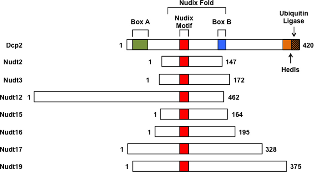
Figure 1. Schematic representation of the Nudix proteins with decapping activity.
Nudix proteins with decapping activity are aligned relative to their Nudix motif. The human Dcp2 protein is schematically depicted at the top with the evolutionarily conserved domains, with Nudix Motif (red), Box A (green), Box B (blue) and regulatory domains encompassing the binding sites for Hedls (orange) and ubiquitin ligases (orange with stripes).

