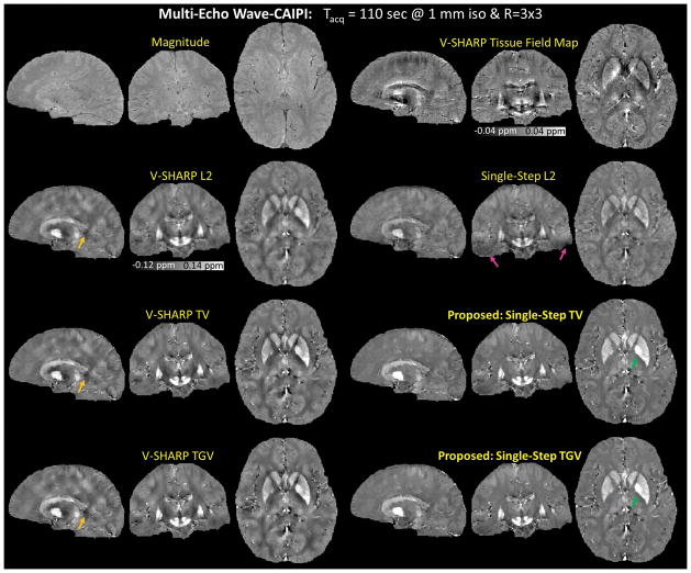
Figure 7.
Multi-Echo Wave-CAIPI. The reconstructed magnetic susceptibility maps obtained from six different QSM algorithms. The proposed methods had the lowest level of dipole artifacts (indicated by the orange arrows), however with reduced contrast between white matter and gray matter (indicated by the green arrows). Compared to Single-Step L2, the proposed methods with the same SMV kernel sizes alleviated the B0 artifacts in the temporal lobes in the Single-Step L2 reconstructed susceptibility map (pink arrows).

