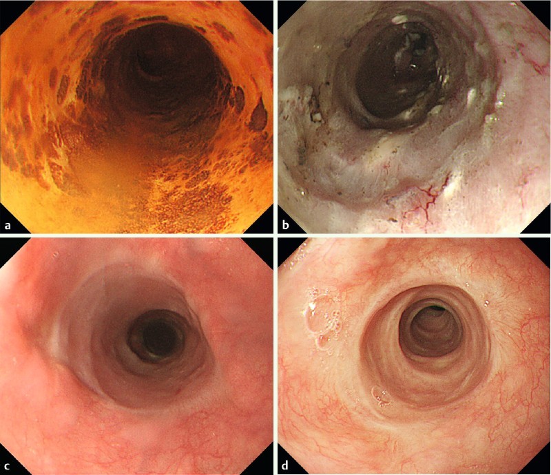
Fig. 3.

Representative case (case 2). 76-year-old male who underwent endoscopic resection for large superficial esophageal squamous cell carcinoma: a Endoscopic view of the tumor after Lugol’s staining. The tumor spread to about 7/8ths of the circumference of the esophageal lumen. b Endoscopic view of the ulcer bed immediately after ESD. The width of the mucosal defect was the entire lumen circumference (Group C). Then, steroid injection followed by oral steroid was administered as a prophylactic treatment. c Endoscopic view on the 35th day. The mucosal defect was still undergoing re-epithelialization, and an ordinary sized endoscope could pass. d Endoscopic view on the 120th day. The complete epithelialization is shown and an ordinary sized endoscope could pass without dysphagia.
