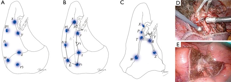
Figure 11.
Segmentectomy completed by three-dimensional stapling. (A,B) The design of three-dimensional stapling in right S9 segmentectomy as an example; (C) completed S9 segmentectomy using stapler-based VAL-MAP-assisted approach. Note that multiple points such as r1–r3 and t1–t3 shown in (B) are stapled together into points R and T. The stapling process in 3D is very difficult to imaging during surgery. However, by simply following the principle including “going peripherally to centrally” “step-by-step stapling” following the “standing” stitch markings based on the geometric information provided by VAL-MAP, such a challenging 3D segmentectomy can be completed under thoracoscopy. The completed figure is typically like that shown in (C); the diaphragm is pulled up close to the hilum (points R and T); (D) an intraoperative view after completion of three-dimensional segmentectomy (S8a + S9 + 10b); (E) an intraoperative view after inflation of the remaining lung in the same case as (D). Once the lung is inflated, the remaining segments are nicely expanded with good post-operative image and function.

