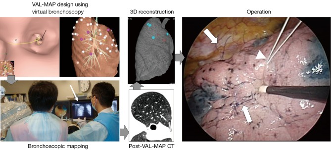
Figure 2.
Steps in VAL-MAP. In general, VAL-MAP and the following surgery are conducted in five steps. Following design of the lung “map” using virtual bronchoscopy, VAL-MAP is usually conducted on the same day or a day before surgery. CT scan after VAL-MAP is taken within 2–3 hours after VAL-MAP, showing actual locations of markings (arrow). Using a radiology workstation, 3D images are reconstructed based on post-VAL-MAP CT scan, reflecting actual locations of markings. The operation field should look the same as that of 3D images. In segmentectomy, the “standing” stiches (arrows) are placed along resection lines, referring to the lung “map” made by VAL-MAP. Another stich indicating the location of the tumor may also be placed using a stich with different color (arrow head). Note that the “standing” stiches are not necessarily placed at the dye markings made by VAL-MAP; dye markings provide geometric information, based on which ideal resection lines are determined and then the resection lines are indicated by the “standing” stiches. VAL-MAP, virtual assisted lung mapping.

