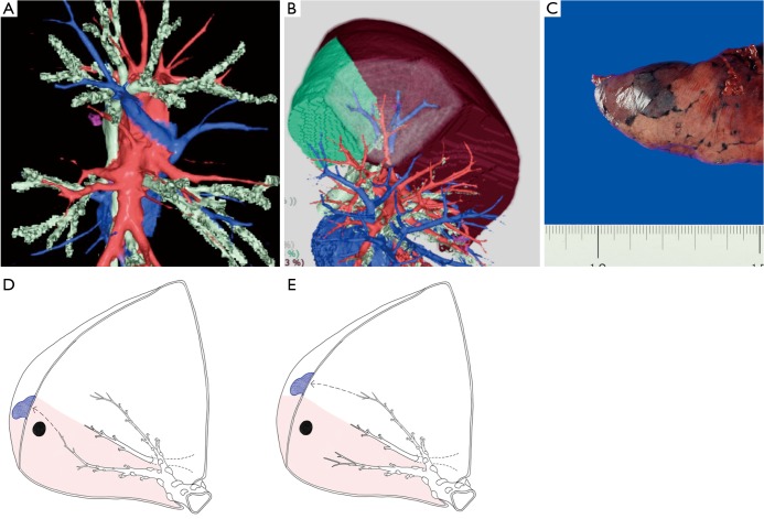
Figure 3.
The principle of VAL-MAP-assisted segmentectomy. (A) A three-dimensional image made by a radiology workstation (Synapse Vincent®) showing hilar vascular structures; (B) a lung resection analysis of Synapse Vincent showing the area of subsegments and VAL-MAP markings in combination with hilar structures; (C) a good marking should not disseminate across the intersegmental plane, staining only a single lobule; (D) a marking is placed close to the intersegmental line from a bronchus inside the target segment (shadowed area); (E) a marking is placed close to the intersegmental line from a bronchus outside the target segment (shadowed area). The panels C, D, and E are reproduced with permission from Cancer Res Front 2016;2:85-104.

