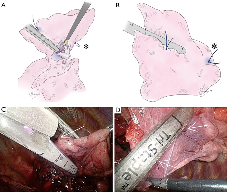
Figure 8.
The principle of the “step-by-step” stapling technique. A stapler should be placed from the periphery to the central area. The position of both the cartilage (A) and anvil (B) of the stapler should be visually confirmed by twisting the lung, changing the angle of the thoracoscope, or changing the camera port. The cartilage rather than the anvil of a stapler should be placed toward the hilum to avoid the risk of vascular damage. Also, the anvil tends to stick into the lung parenchyma, causing bleeding. The “standing” stitch markings should be used as a guide to determine the location of stapling and to determine the direction. Note that the most cephalic stitch (*) can be seen from both sides because it is standing out of the lung; (C,D) intraoperative views corresponding to (A) and (B). Arrows indicate “standing” stiches.

