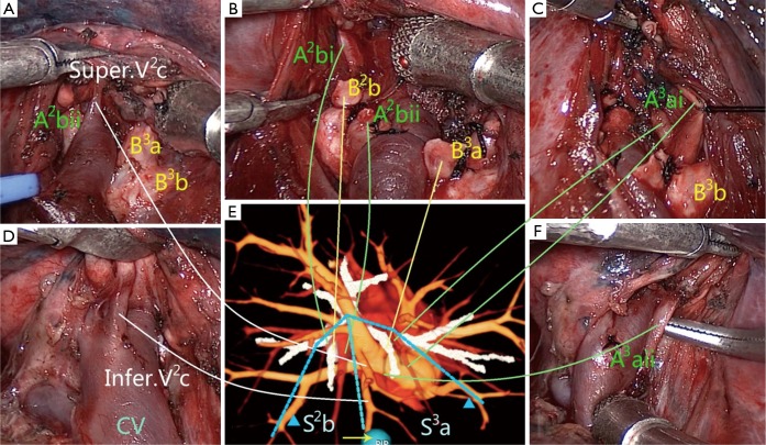
Figure 2.
Illustration of the surgical procedure with the assistance of 3D image navigation. The sequence of the surgical procedure was from (A-D,F). The dissection begun at the central vein, and all following dissections were kept going along it. (E) 3D image from the right superior view revealed the anatomic structures of the right upper lobe. The targeted subsegments were S2b and S3a, the targeted arteries were A2bi, A2bii, A3ai and A3aii, the targeted bronchi were B2b and B3a, and the targeted veins were inferior V2c and superior V2c. The veins marked with blue triangles were the intersubsegmental veins, which were the marks of intersubsegmental demarcation and should be preserved. All solid lines reflected the one-to-one correspondence between the actual and virtual anatomic structures.

