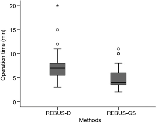
Figure 3.

The operation time after visualization of PPLs for REBUS-GS-TBB and REBUS-D-TBB (P=0.00053). PPLs, peripheral pulmonary lesions; REBUS-GS, radial endobronchial ultrasound with a guide sheath; REBUS-D, REBUS with distance; TBB, transbronchial biopsy.
