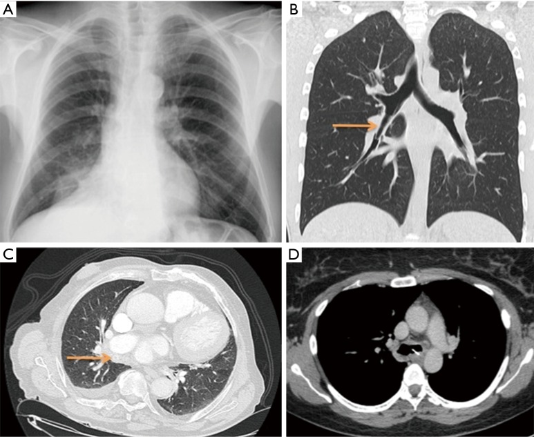
Figure 1.
Radiological images showing complications and/or different radiopaque foreign bodies. (A) Posteroanterior chest radiograph. We can observe a consolidation in the medium lobe caused by chicken bone; (B) coronal image of CT. Arrow is showing a Bic cap lodged in the bronchus intermedius; (C) axial image of CT. Right pleural effusion and a cherry pit indicated by arrow; (D) axial image of CT. We can observe a deer bone lodged in left main bronchus. CT, computed tomography.

