Figure 3.
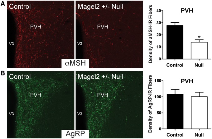
Altered aMSH neural projections to the PVH in Magel2-null mice. Confocal images and quantification of (A) aMSH and (B) AgRP immunoreactive fibers innervating the PVH in adult (P60) Magel2-null and control mice (n = 4–5 per group). V3, third ventricle. Values are shown as the mean ± SEM. *P < 0.05 versus the control.

