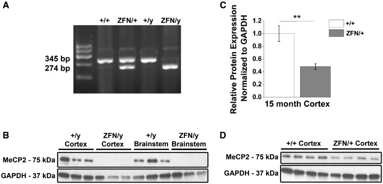
Figure 1.
Mecp2ZFN/y male and Mecp2ZFN/+ female rats display altered MeCP2 protein expression. (A) Representative genotyping gel revealing DNA fragments of different lengths that differentiate WT from Mecp2ZFN/y and Mecp2ZFN/+ rats. (B) Western blotting demonstrates total absence of MeCP2 protein in both cortex and brainstem of PND 60–80 Mecp2ZFN/y rats. (C–D) 15-month-old Mecp2ZFN/+ rats show a ∼52% reduction in MeCP2 protein by western blotting of cortical tissue (WT n = 4 Mecp2ZFN/+ n = 4). Data are presented as mean ± SE, with asterisks representing significant genotype differences (**P < 0.01).

