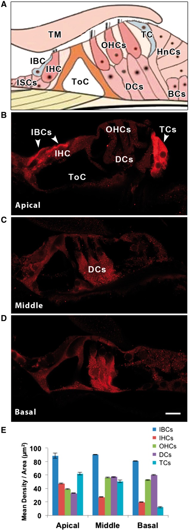
Figure 2.

(A) Schematic diagram of the organ of Corti to show the location of tectal cells (TCs) and inner border cells (IBCs). PKCB II immuno-labelling in the three turns of the rat cochlea, apical (B), middle (C) and basal (D). Cell types are labelled in A, B and C. Note that the apical IBCs and TCs are most intensely labelled, followed by basal and middle turn Deiter’s cells (DCs). Hair cells, both inner (IHCs) and outer (OHCs), are barely above background level. Other abbreviations: tectorial membrane (TM), inner sulcus cells (ISCs), tunnel of Corti (ToC), Hensen’s cells (HnCs), Boettcher’s cells (BCs). (E) Mean density per area of PKCB II for each cochlear cell type and turn of the cochlea. Scale bar in D = 10 µm, applies to B-D.
