Figure 3.

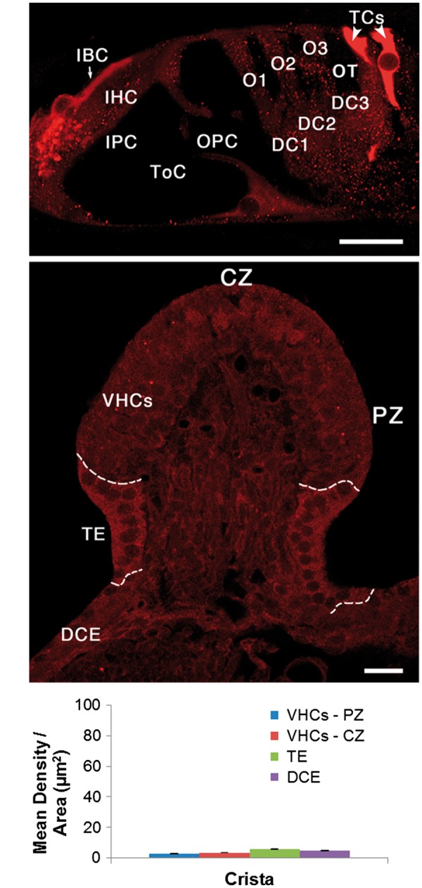
Here we directly compare cochlear PKCB II immuno-labelling in the apical turn of (A) the rat cochlea with (B) the vestibular crista ampullaris in the same animal. Note that the entire vestibular epithelium is less intensely labelled than the cochlear inner border cells (IBCs) and tectal cells (TCs). Dashed lines in B separate the vestibular sensory epithelium (VHCs) from the transitional epithelium (TE), and the TE from the dark cell epithelium (DCE). (C) Mean density per area of PKCB II in different regions of the rat crista ampullaris. Note this histogram is the same scale as that in Fig. 2E. Other abbreviations: IHC, inner hair cell; IPC, inner pillar cell; ToC, tunnel of Corti; OPC, outer pillar cell; O1, O2, O3, first, second and third row outer hair cells; DC1, DC2, DC3, first, second and third row Deiters cells; OT, outer tunnel. CZ, central zone; PZ, peripheral zone; VHCs, vestibular hair cells (sensory epithelium), TE transitional epithelium (supporting cells). Scale bars = 10 µm.
