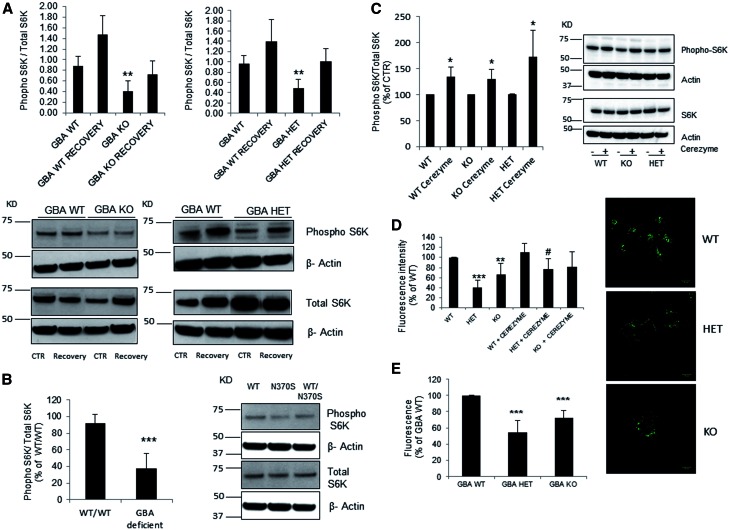
Figure 2.
Impairment of autophagic lysosome reformation in GCase deficient cells. (A) Gba1 KO and Gba1 HET presented decreased basal levels of phopho-S6K compared to Gba1 WT (n = 4). The percentage of recovery was similar between all cell lines (40-50% increase compared with basal) however the levels of phopho-S6K were consistently lower in Gba1 KO and Gba1 HET cells. (B) Phopho-S6K protein levels were significantly lower in mutant GBA1 patient derived fibroblasts (n = 5) compared to WT fibroblasts. (C) Phospho-S6K levels were significantly increased in WT, HET, KO Gba1 MEF cells upon cerezyme treatment (n = 4). Results were expressed as % of respective control. (D) Lyso ID fluorescence measurements detected a significant decrease in acidic functional lysosomes in both Gba1 HET and Gba1 KO compared to Gba1 WT (n = 4). Cerezyme treatment significantly increased acidic functional lysosomes in Gba1 HET MEFs but not Gba1 KO. Results were expressed as % of respective control. #P < 0.01 for HET + cerezyme compared to HET. Scale bar 10µm. (E) Lyso ID fluorescence measurements using a plate reader detected a significant decrease in functional/acidic lysosomes in both Gba1 HET and Gba1 KO compared to Gba1 WT (n = 4). All data represent mean ± SD, *P < 0.05; **P < 0.01, ***P < 0.001 vs. respective control.

