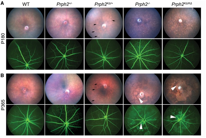
Figure 4.
K153Δ leads to fundus flecking characteristic of PD. (A–B) Funduscopic examination was performed at the indicated genotype at P180 (A) and at P365 (B). Shown are representative brightfield fundus images (top) and fluorescein angiograms (bottom). Arrows indicate flecking phenotype found in the Prph2KΔ/+ as well as the Prph2-/- and Prph2KΔ/KΔ. Arrowheads indicate splotching, likely due to severe photoreceptor degeneration, which occurs at later ages. n = 6-8 eyes/group.

