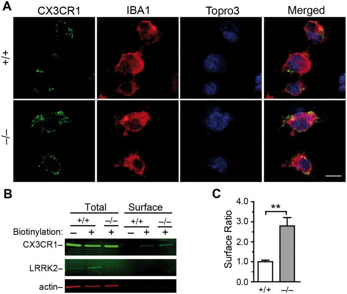
Figure 3.
Lrrk2–null microglia show increased presentation of CX3CR1 at the cell surface. (A) Representative images show CX3CR1 staining of impermeabilized microglia. The cells were counterstained with IBA1 and Topro3. Scale bar, 10 μm. (B) Western blot analyses the levels of CX3CR1. LRRK2 and actin at cell surface and in total lysates. (C) Bar graph depicts that the ratio of CX3CR1 proteins at cell surface in cultured Lrrk2+/+ and Lrrk2–/– microglia. Values represent means ± SEM. Significant differences between the groups are expressed as follows: **P < 0.01. The experiments were repeated three times independently.

