
Figure 5.
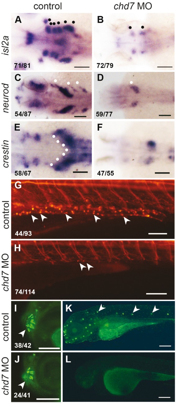
chd7 knockdown causes loss of peripheral neuronal lineages. (A, B) RNA in situ hybridization of islet 2a at 72hpf which marks the differentiated sensory and motor cranial neurons (black dots, a) was severely reduced in chd7 morphants. (C, D) The epibranchial neuronal precursors marked by neuroD in the head (white dots, C) was lacking in the chd7 morphants. (E, F) The vagal neural crest and precursors of enteric neurons marked by crestin in 36hpf control embryos (white dots, E) was reduced in chd7 morphants. (G,H) At 6dpf, Tg(NBT:dsRed) marks enteric neurons on the gut tube (white arrowheads, g) was severely decreased in chd7 morphants. (I, J) Tg(sox10:eGFP) marks well patterned and organised microvillous neurons at 4dpf (white arrowheads, i) and fewer and disorganized microvillous neurons were present in chd7 morphants. (K,L) DASPIE staining highlights the neuromasts cells along lateral line in 4dpf control embryos (white arrowheads, k) and these neurons were completely absent in the chd7 morphant embryos. All images have anterior to the left, dorsal views (A–F) and lateral views (G–L). Scale bars are 100µm (A–H, K–L) and 50µm (I–J). The numbers in the bottom left corner indicate the actual number of embryos of the total represented by the image.