Figure 6.
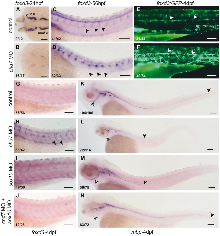
chd7 knockdown leads to expanded non-myelinating glia and reduced myelinating Schwann cells in a Sox10-dependent manner. (A, B) Cranial glia associated with pre-otic and post-otic ganglia in 24hpf embryos was completely absent in the chd7 morphant embryos. (C, D) In 56hpf embryos foxd3 positive satellite glia associated with the dorsal root ganglia are enhanced in the chd7 morphants. (E, F) Tg(foxd3:GFP) marks the lateral line glia in 4dpf control larvae (arrowheads, E), which shows increased fluorescence intensity with many more dentritic shaped cells present ectopically in the chd7 morphant embryos (injected with 0.8ng of chd7 MO) (arrowheads, F). (G–J) foxd3 RNA in situ hybridization of 4dpf larvae does not show any signal in control, while chd7 morphant embryos have enhanced signal. sox10 morphants and embryos coinjected with chd7 and sox10 MO are not different from the control. (K–N) RNA in situ hybridization for myelin basic protein (mbp) in 4dpf embryo marks the myelinated glia. The extend of myelinating Schwann cells on the lateral line are marked by the black arrowhead and the periocular ganglia are marked by black open arrowheads. .The chd7 morphant embryos (L) and the sox10 morphant embryos (N) have reduced extend of lateral line glia (black arrowhead) and periocular glia (open arrowhead). Emrbyos coinjected with chd7 MO and sox10 MO show a recovery in the posterior extend of the lateral line Schwann cells (black arrowheads N). All images have anterior to the left, dorsal views (A–B) and lateral views (C–N). All scale bars are 100µm. The numbers in the bottom left corner indicate the actual number of embryos of the total represented by the image.

