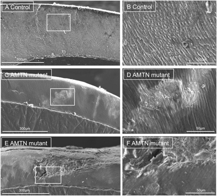
Figure 4.
SEM of representative exfoliated teeth. (A and B) SEM of tooth 1 from the control individual, (C–F) SEM of tooth 4 from individual IV:1, boxed regions on pictures A, C and E reflect the boundaries of the photographs taken at higher power in these regions, labelled B, D and F. Control tooth 1 exhibits normal, typical enamel architecture comprising prisms (rods) of individual enamel crystallites. Tooth 4 exhibits both regions of relatively normal enamel and disturbed structure. The cross sectional surface of Tooth 4 has a “smooth” appearance that may reflect the presence of organic material.

